Expert Review: This article includes clinical insights from Dr. Vivek Arora, a physical therapist (physiotherapist) with 20+ years of experience.
If you have just read a radiology report and started searching bulging disc vs herniated disc, you are asking a reasonable question with an inconvenient answer. Yes, the two terms mean genuinely different things. No, the difference is not the one most people assume. A bulge and a herniation are descriptions of shape — the outline of disc tissue on a scan, measured against the edge of the vertebral bone. They are not severity ratings, they are not diagnoses, and on their own they say remarkably little about how much your back or leg will hurt next Tuesday.
That gap between what the words sound like and what they actually mean causes a lot of unnecessary worry. “Bulging” sounds mild and reassuring. “Herniated” sounds like something tore and is now loose inside you. In practice, people walk around with bulges and feel nothing at all, while a small, technically less dramatic herniation sitting in exactly the wrong spot can produce leg pain severe enough to keep someone off work. This article explains the vocabulary properly, then explains why the vocabulary matters far less than what your symptoms are doing.
Key takeaways
- The difference is real but geometric. A bulge is broad and generalized, involving more than a quarter of the disc’s circumference. A herniation is focal, involving less than a quarter.
- Bulges are extremely common in people with no pain at all. Their prevalence rises steadily with age, which is part of why the finding alone explains so little.
- Herniations subdivide further into protrusion, extrusion and sequestration — again describing shape, not damage severity or prognosis.
- What decides your symptoms is nerve contact and available space, not the label. Position relative to the nerve root, inflammation, and how much room the canal had to begin with matter far more.
- Initial management is essentially identical for both. Neither word, by itself, is a reason for surgery or a reason to stop moving.
What is a spinal disc actually doing when it bulges or herniates?
A spinal disc is a pressurized spacer between two vertebrae. It has a firm outer wall of layered fibres called the annulus fibrosus, and a softer, water-rich centre called the nucleus pulposus. Bulging means the whole outer wall has spread outward beyond the bony rim, like an under-inflated tyre spreading under load. Herniation means inner material has pushed out through a localized weakness in that wall.
The mechanics are worth understanding because they explain why the two shapes form differently. The annulus is built in crossing layers, a bit like plywood, which makes it excellent at resisting the twisting and shearing forces of everyday movement. The nucleus behaves more like a gel that redistributes pressure whenever you bend, lift or sit. When you flex forward, the nucleus loads the back wall of the disc more heavily — which is exactly why the back of the disc, nearest the nerves, is where most problems appear.
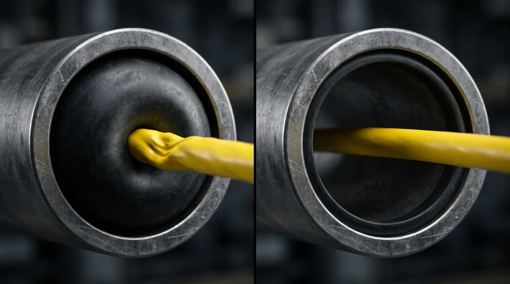
Over decades, discs lose water content and height. The annulus becomes less springy and spreads outward around most of its rim. That is a bulge, and it is largely a maturation change rather than an injury. A herniation is a different mechanical event: pressure finds a weak point in the annular layers and drives nucleus material through it in one focused direction. That can happen suddenly with a lift or a twist, but it far more often builds over weeks of accumulated loading before one ordinary movement becomes the last straw. If you have ever noticed that a flare-up starts after bending to pick up something trivial, that is the pattern — the sock was not the cause, it was the timing.
Bulging disc vs herniated disc: what your MRI report is actually describing
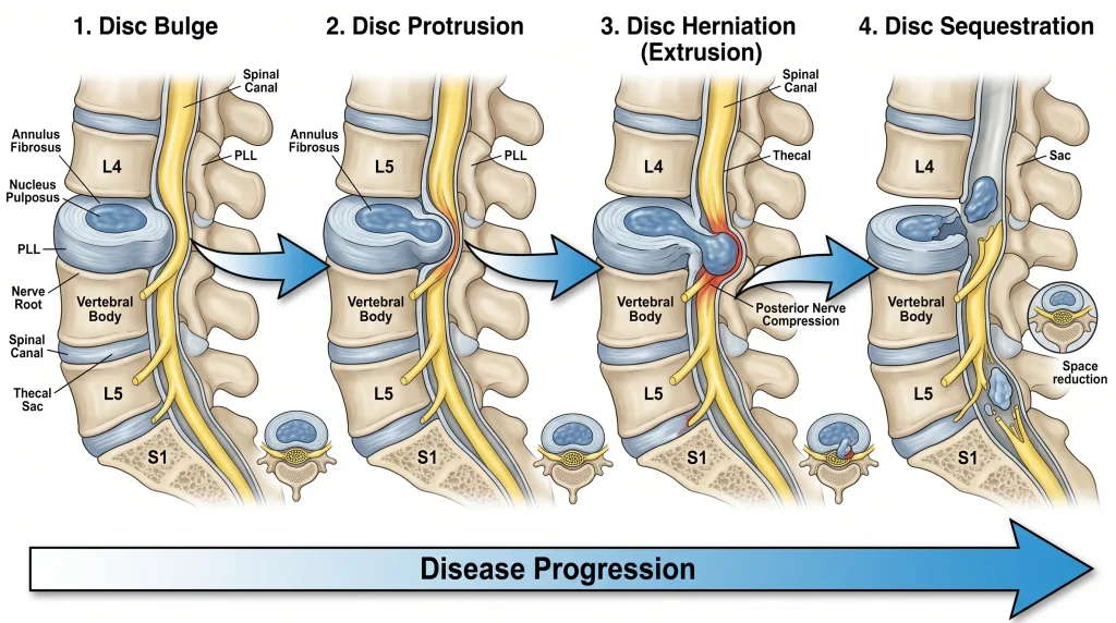
Radiologists use a standardized vocabulary agreed by the North American Spine Society, the American Society of Spine Radiology and the American Society of Neuroradiology. In that system, a bulge is generalized displacement of disc tissue beyond the vertebral margins involving more than 25% of the disc’s circumference. A herniation is localized displacement involving less than 25%. That percentage is the entire distinction. It is a measurement of how much of the rim is involved, not of how bad things are.
Herniations are then described further by their profile. A protrusion is a herniation where the base — the neck of tissue still connected to the disc — is wider than the part sticking out. An extrusion is the reverse: the displaced portion is wider than its base, meaning material has pushed through the annular fibres rather than simply stretching them. A sequestration is a free fragment that has separated from the parent disc entirely. You may also see the phrase broad-based, which sits in the overlap zone between focal and generalized. And you will almost certainly see central, paracentral, foraminal or extraforaminal — these describe where around the circle the material sits, and clinically they are often the most informative words in the whole report.
| Term on the report | What it means anatomically | How often it appears in people with no pain | What it predicts about symptoms | What it changes about early treatment |
|---|---|---|---|---|
| Disc bulge | Generalized outward spread of the disc wall past the bony rim, involving more than 25% of the circumference | Very common and rises with age — roughly a third of pain-free 20-year-olds and the majority of pain-free 50-year-olds | Very little on its own; frequently an incidental finding | Usually nothing |
| Disc herniation (umbrella term) | Focal displacement of disc material involving less than 25% of the circumference | Common as protrusions; less common in more advanced forms | Depends almost entirely on position and nerve contact | Usually nothing at the initial stage |
| Protrusion | Herniation whose base is wider than the displaced portion — the annular fibres are stretched, not breached | Present in roughly a third of pain-free adults across most adult ages | Weak predictor; a small one in a tight recess can still hurt a great deal | Nothing by itself |
| Extrusion | Displaced portion is wider than its base — material has pushed through the annular fibres | Considerably less common as an incidental finding | More often associated with nerve root symptoms, but also more likely to shrink over time | Nothing by itself; may raise interest if leg symptoms match the level |
| Sequestration | A free fragment separated from the parent disc | Uncommon incidentally | Can produce marked radicular pain — and free fragments are among the most likely to resorb | Still not an automatic surgical indication |
This is the same interpretive trap that catches people reading any spinal imaging report. Plain films generate their own vocabulary of alarming-sounding but usually unremarkable phrases, which is why a plain-language guide to what X-ray report wording actually means is often more reassuring than the report itself. The MRI equivalents behave the same way: Modic changes in the vertebral endplates and mild canal narrowing described as stenosis both sound far more decisive on paper than they usually prove to be in the clinic.
Why doesn’t the label predict how much it hurts?
Because the label describes the disc, and your symptoms are usually produced by the nerve. Pain, numbness and weakness in a leg come from a nerve root being mechanically compressed, chemically irritated, or both. Whether that happens depends on where the displaced material sits relative to the root, how much spare room the canal and lateral recess had before anything moved, and how inflamed the local tissue is. Shape alone answers none of those questions.
The prevalence data makes the point bluntly. In a systematic review of imaging findings in people with no back symptoms at all, disc bulges appeared in about 30% of 20-year-olds and around 60% of 50-year-olds, while disc protrusions were present in roughly 29% to 43% of pain-free adults across the age range (Brinjikji et al., 2015). If a finding is that common in people who feel fine, it cannot be doing much explanatory work on its own.
Three factors do far more of the work:
- Position. A paracentral protrusion sitting directly in the lateral recess — the narrow gutter where a nerve root turns to exit — can be small and still be miserable. The same volume of tissue displaced centrally, where there is more room, may cause nothing.
- Available space. Canal and foraminal dimensions vary a lot between people. Someone with a naturally roomy canal absorbs a herniation without symptoms; someone with a congenitally narrow one has no tolerance to spare.
- Inflammation. Nucleus material that reaches the epidural space is chemically irritating to nerve tissue. This is part of why symptoms can be severe without dramatic compression — and part of why they often settle substantially before any imaging change occurs.
There is a further irony that surprises most patients. Larger extrusions and free fragments are, on average, more likely to shrink over time than small contained bulges are. Material that has breached the annulus is exposed to the body’s immune and vascular response, which can gradually break it down. A broad degenerative bulge, by contrast, is a structural change in the disc wall and is not going anywhere. The scarier-sounding finding often has the better natural history.
How do I tell whether a nerve is actually involved?
By what the symptoms do, not by what the report says. Nerve root involvement typically produces pain that travels in a defined band down the leg rather than spreading vaguely, often below the knee, and frequently with pins and needles, numbness or weakness in a matching distribution. Purely local back pain that stays around the belt line — even with an impressive-looking scan — usually points elsewhere.
A few patterns are worth recognizing. Radicular pain tends to be sharp, electrical or burning rather than dull and aching, and it usually has a consistent route. The specific sensations that identify true nerve root pain are surprisingly reproducible from person to person. Symptoms that spike momentarily with a cough, sneeze or bearing down are also informative, because those actions briefly raise pressure inside the spinal canal — a sharp jolt with sneezing is one of the more disc-suggestive things a person can report.
What makes it better and worse matters too. Disc-related symptoms commonly worsen with sustained sitting, forward bending and the first movements after a long car journey, and ease with standing, walking or lying flat. Symptoms that ease specifically when you lean forward and worsen when you stand upright or walk downhill point more toward canal narrowing than toward a fresh disc problem. And pain that is worse in the second half of the night with morning stiffness lasting well over half an hour raises the question of an inflammatory cause rather than a mechanical one — a different conversation entirely.
Quick check
Five yes/no questions that separate “an interesting scan finding” from “a nerve that needs attention”:
- Does the pain travel below the knee in a consistent band, rather than spreading vaguely?
- Is there numbness, pins and needles, or a patch of altered sensation in the same route?
- Can you feel a definite jolt down the leg when you cough or sneeze?
- Is there any genuine weakness — catching your toe on steps, difficulty rising onto the ball of one foot, a foot that slaps?
- Has the leg pain now become clearly worse than the back pain?
Two or more yes answers suggests a nerve root is involved and that clinical assessment is worth arranging. Weakness on question four should be assessed promptly rather than monitored at home.
When is a disc problem an emergency?
Rarely — but the exceptions are genuine and time-sensitive. A small number of presentations need same-day medical assessment regardless of what any previous scan said. These are not reasons for general alarm; they are a short list of specific patterns that change the urgency of the situation, and each one is on the list for a concrete anatomical reason.
- New difficulty passing urine, loss of bladder or bowel control, or numbness around the saddle area (inner thighs, genitals, buttocks). These suggest compression of the bundle of nerve roots at the base of the spinal cord. This is a surgical emergency because the window in which function can be preserved is measured in hours to a small number of days, not weeks.
- Weakness in both legs, or weakness that is clearly progressing day by day. Progressive motor loss indicates the nerve is not merely irritated but functionally failing, and the trajectory matters more than the current severity.

- Fever, unexplained weight loss, night pain that will not settle in any position, or a history of cancer. Discs do not cause these. This combination raises the possibility of infection or a non-mechanical cause, which requires a different diagnostic pathway entirely.
- Significant trauma, or any new back pain after a fall in someone with osteoporosis or on long-term steroids. The concern here is vertebral fracture rather than disc displacement, and fractures need to be identified before rehabilitation begins.
- A recent onset of intravenous drug use, or a recent spinal injection or procedure followed by escalating pain. These raise the index of suspicion for spinal infection, which can be indolent early and serious late.
When is imaging actually worth doing?
When the result will change what happens next. That is the whole test. National guidance in the UK advises against routine imaging for low back pain outside specialist settings, and recommends it only where the result would alter management (NICE NG59). For most people in the first few weeks, an MRI will find something — and that something will usually be a bulge or protrusion that a third of pain-free people also have.
Imaging genuinely earns its place in a few situations: when red flag features are present, when leg symptoms are severe or persistent and someone is being considered for an injection or surgery, or when a clinician needs to confirm that a specific nerve root at a specific level matches specific symptoms before intervening. In those settings the scan is answering a question that has already been framed clinically.
What imaging does not do is grade your problem or predict your recovery. It also carries a real cost that gets underestimated: reading the words “extrusion” or “degenerative disc disease” on your own report changes how you move. People who believe their spine is structurally damaged tend to guard, brace and avoid — and guarded, braced, avoidant movement is itself a driver of persistent pain. Scans do not cause that. Uninterpreted scans do.
Three things a disc report does not mean
- “Bulging” does not mean early-stage herniation. They are not points on the same timeline. A bulge is a broad structural change; a herniation is a focal displacement. One does not routinely progress into the other.
- “Herniated” does not mean the disc has slipped. Discs are anchored to the vertebrae above and below. Nothing slides out of place. The material displaces through a wall — which is why the popular phrase is anatomically misleading.
- Neither term means you should stop moving. Prolonged rest reliably makes disc-related pain harder to recover from, not easier.
From the Clinic: Dr. Arora’s Expert Insight
Almost everyone who brings me a disc report has already compared it to someone else’s. There is a colleague who had “exactly the same thing” and was back on the golf course in a month, or a brother-in-law with an identical-sounding scan who ended up in an operating theatre and still limps. The comparison arrives before the history does, and it is usually doing a lot of quiet emotional work — either setting up a timeline the person expects to meet, or a fate they are bracing for. So the first thing I do is take the comparison seriously rather than dismiss it, and then ask what their colleague’s leg was doing. Nine times out of ten they do not know. That is the point I want to land: two people can share a word on a report and have completely different problems, because the word describes the disc and the problem lives at the nerve.
What often gets missed in that comparison is directional preference — how symptoms respond to repeated movement in one direction versus the other. It is one of the few things I can test in a few minutes that actually changes what I prescribe that afternoon. Two people with the same reported protrusion at the same level can respond in opposite ways: one centralizes their leg pain with repeated extension and does beautifully with a loading strategy built around it, while the other flares with the same movement and needs an offloading and flexion-biased approach first. Nothing on the MRI tells me which person is in front of me. Only the movement response does. This is also why forum advice about “the exercises for a herniated disc” is such a coin flip — half the readers are being handed the wrong direction.
The place generic advice fails hardest with disc problems specifically is the sitting instruction. “Avoid prolonged sitting” is technically correct and practically useless, because it gives no threshold and no alternative. What works better is finding the person’s actual sitting tolerance — often eight to twelve minutes early on, which people find shocking — and then building a break rhythm just inside it, rather than sitting until it hurts and then standing up. Interrupting the load before symptoms escalate produces far better weeks than reacting to symptoms after they arrive. The same logic applies to driving, which is the single most common reason a well-managed disc flares in week three of an otherwise good recovery.
What should you do in the first 72 hours?
Keep moving in small, frequent, tolerable doses, protect the positions that provoke symptoms, and stop trying to establish which disc is responsible. Early management for a bulge and for a herniation is the same, because in both cases you are managing an irritated nerve and a sensitized back, not a shape on a scan. The immediate goal is to reduce provocation enough that normal movement becomes possible again.
What to do today
- Walk in short, frequent bouts — five to ten minutes at a time, several times a day, rather than one long attempt. Flat ground, unhurried pace.
- Cap your sitting before it bites. Find the number of minutes you can sit comfortably, subtract two, and stand up at that point every time.
- Change how you get out of bed and out of the car. Roll to your side and push up with your arms; swing both legs out together before standing. Twisting under load through a fresh disc irritation is the most reliable way to lose a day.
- Postpone deep forward bending, deadlifts, rowing machines and sit-ups for now — not forever, and not because bending is dangerous, but because early flexion under load is the position that most reliably provokes a fresh disc problem.
- Track the leg, not the back. If the leg symptoms are retreating upward toward the buttock over days, you are heading in the right direction even if the back still aches.
How does rehab progress from there?
In stages defined by what your symptoms are doing, not by the calendar and not by the wording of your scan. Progression is earned by two things: the leg symptoms retreating toward the spine, and your tolerance for sitting, walking and bending increasing without a next-day payback. Advancing on a fixed timetable while symptoms are still travelling further down the leg is the most common way rehab goes backwards.
| Stage | Main goal | What it typically involves | Ready to progress when |
|---|---|---|---|
| 1. Settle and desensitize | Reduce nerve irritation; restore basic daily movement | Frequent short walks; position management; gentle movement in the direction that eases symptoms; nerve gliding if tolerated | Leg symptoms have retreated above the knee, or clearly reduced in intensity; you can sit 20 minutes without a next-day increase |
| 2. Restore hip and trunk control | Get load off the lumbar spine by making the hips do their share | Hip hinge practice with a dowel; glute bridges and progressions; side-lying hip abduction; controlled dead bug variations | You can hinge to mid-shin without symptoms travelling down; walking 30 minutes is comfortable; no morning symptom spike after activity |
| 3. Load and build tolerance | Rebuild capacity so ordinary loading stops being a threat | Goblet squats, light Romanian deadlifts, split squats, farmer’s carries, gradual return to gym or sport-specific patterns | Full pain-free range in daily tasks; loaded hinge with 20–30 kg (45–65 lb) with no leg symptoms; strength symmetrical between sides |
| 4. Return and protect | Return to full activity and reduce recurrence risk | Progressive return to previous loads and volumes; maintaining two strength sessions weekly; managing sitting and driving exposure | Ongoing — this stage does not end, it becomes the maintenance habit |
Exercise selection matters more than exercise volume here. Hip hinge work is central because a stiff, under-used hip forces the lumbar spine to absorb bending load it was not designed to take repeatedly. Posterior chain strength changes the distribution of that load rather than merely “strengthening the core.” If you want a structured starting point, a progressive home physical therapy program for the lower back gives a reasonable framework to work from — though the direction of loading should be matched to how your symptoms actually respond, not chosen from a list.
Do’s and don’ts for a disc-related problem specifically
- Do break up sitting proactively, before symptoms arrive. Reactive standing is too late.
- Do use the leg symptom’s position as your primary progress marker — retreating upward is good, travelling downward is a signal to back off.
- Do get the seat position sorted in your car early, since driving combines sitting, vibration and a twisted exit.
- Don’t repeatedly test the painful movement to see if it still hurts. Repeated provocation keeps the nerve sensitized.
- Don’t stretch aggressively into the leg pain. Hamstring stretching in particular often puts tension on an already irritated nerve rather than a tight muscle.
- Don’t chase repeat imaging to see whether the herniation has shrunk. Imaging change and symptom change run on different clocks, and the scan will not tell you what to do next.
What if conservative care stalls, and when does surgery become relevant?
Conservative care is considered stalled when six to twelve weeks of genuinely structured rehabilitation produces no meaningful change in leg symptoms or function — not when pain fluctuates, and not when a scan looks unchanged. At that point the useful next steps are a reassessment of the working diagnosis, consideration of an image-guided injection to reduce nerve inflammation, or specialist referral. Surgery enters the conversation only in specific circumstances.
Before assuming the rehab failed, it is worth checking that it was actually the right rehab. Stalled cases frequently turn out to be under-dosed, mis-directed, or aimed at the wrong structure entirely — hip-origin pain, a sacroiliac contribution, or an inflammatory pattern hiding behind a mechanical story. A plateau is a prompt to re-examine, not automatically to escalate.
When surgery is considered, the decision rests on two things aligning: non-surgical treatment has not improved pain or function, and the radiological findings genuinely match the symptoms and clinical signs (NICE NG59). That second condition is where the bulge-versus-herniation question finally acquires some practical weight — a broad degenerative bulge with no root correlation is a poor surgical target, whereas an extrusion sitting on the exact root that explains a person’s weakness and dermatomal pain is a coherent one. The exception is the emergency presentation described earlier, where cauda equina compression or rapidly progressing weakness moves surgery from an elective decision to an urgent one.
The realistic outlook is better than most people expect. The majority of disc-related leg pain improves substantially over weeks to a few months with structured conservative management, and a meaningful proportion of herniated material reduces in size over time without any intervention. Recurrence is possible, which is why the strength and load-management habits built in the final stage are worth keeping rather than abandoning once the pain has gone.
The bottom line
In the bulging disc vs herniated disc question, the honest answer is that there is a real difference and it is smaller than it sounds. A bulge is broad and generalized; a herniation is focal and subdivides into protrusion, extrusion and sequestration. Both are descriptions of shape on an image. Neither is a diagnosis, a severity score, or an instruction. What determines how you feel is whether displaced material is contacting or inflaming a nerve root, where it sits, and how much room there was to begin with.
So do this instead of re-reading the report: for the next two weeks, write down two numbers each evening — how far down your leg the symptoms travelled at their worst, and how many minutes you could sit comfortably. If the first number is climbing back up your leg and the second is growing, your rehab is working and the wording on the scan is irrelevant. If neither number is moving after three or four weeks of consistent effort, that is your cue to get reassessed — and it is a far more useful trigger than any noun a radiologist chose.
Frequently asked questions
Is a bulging disc less serious than a herniated disc?
Not reliably. A bulge involves more of the disc’s circumference but is often an age-related change with no symptoms at all, while a small herniation in a tight space can cause severe leg pain. Seriousness is determined by nerve involvement and functional impact, not by which of the two words appears on the report.
Can a bulging disc turn into a herniated disc?
It can, but they are not stages of the same process. A bulge is a broad structural change in the disc wall, while a herniation is a focal breach at one weak point — a disc can have both, or either, independently. Having a bulge on a scan does not mean a herniation is coming.
Does herniated disc material ever go away on its own?
Yes, and often more than people expect — extrusions and free fragments in particular can reduce in size over months as the body breaks the material down. Broad degenerative bulges generally do not resorb in the same way. Symptom improvement, however, usually arrives well before any imaging change.
Will a bulging or herniated disc show up on an X-ray?
No — X-rays image bone, not disc tissue, so a bulge or herniation cannot be seen directly. What an X-ray may show is reduced disc height, which suggests degenerative change but says nothing about displacement or nerve contact. MRI is the modality that visualizes disc material.
Can I still go to the gym with a disc finding on my scan?
In most cases yes, with the loading pattern adjusted rather than the gym abandoned. Deep loaded flexion — conventional deadlifts, rowing machines, weighted sit-ups — is usually the first thing to modify, while carries, split squats and hip-dominant work often remain well tolerated. Leg symptoms travelling further down during or after a session is the signal to reduce load.
Does a bigger herniation mean I am more likely to need surgery?
Not on size alone. Surgical decisions rest on persistent disabling symptoms after adequate conservative care combined with imaging that matches those symptoms, or on emergency features such as cauda equina signs. Some of the largest herniations resolve without any procedure at all.
Should I ask for an MRI if I have not had one?
Only if the result would realistically change your treatment in the next few weeks. Without red flag features or severe persistent leg symptoms under specialist review, an early MRI usually finds common age-related changes and adds worry rather than direction. If your symptoms are settling with structured rehabilitation, a scan changes nothing.
Medical disclaimer
This article explains radiological terminology and general conservative management principles for lumbar disc findings. It cannot interpret your individual scan, and no article can — a report’s meaning depends on your symptoms, your neurological examination and your history, none of which are visible here. If you have leg weakness, changes in bladder or bowel function, saddle numbness, fever, unexplained weight loss, or a history of cancer alongside your back pain, seek medical assessment promptly rather than relying on this or any other online explanation. Nothing here replaces an individual assessment by a qualified clinician.
References
- Brinjikji W, Luetmer PH, Comstock B, Bresnahan BW, Chen LE, Deyo RA, et al. Systematic literature review of imaging features of spinal degeneration in asymptomatic populations. American Journal of Neuroradiology. 2015;36(4):811–816. https://doi.org/10.3174/ajnr.A4173
- National Institute for Health and Care Excellence. Low back pain and sciatica in over 16s: assessment and management (NG59). Published 2016, last updated 2020. https://www.nice.org.uk/guidance/ng59