Expert Review: This article includes clinical insights from Dr. Vivek Arora, a physical therapist (physiotherapist) with 20+ years of experience.
A radiology report can do a remarkable amount of damage in about eleven words. Extrusion. Impingement. Nerve root compression. Read on a phone screen in a parking lot, that language sounds structural and permanent, and the question that follows is almost always the right one: can a herniated disc heal on its own?
The short answer is yes, and it happens more often and more completely than most people expect. The longer answer is where the real reassurance lives, because the body’s method for clearing displaced disc material is genuinely counterintuitive — and it explains why the scans that read worst on paper are frequently the ones that improve most.
Key takeaways
- Most lumbar disc herniations shrink without surgery. The immune system recognises displaced disc material as foreign and progressively breaks it down over weeks to months.
- Larger herniations tend to resorb more readily than small contained bulges. More exposed material means more surface for the immune response to work on.
- “Healed” means a pain-free, load-tolerant back — not a pristine MRI. The annular tear repairs with fibrous scar tissue, and lost disc height generally does not come back.
- Pain, numbness and weakness recover on three different clocks. Pain usually improves first and fastest; sensory and motor changes follow a slower and less complete curve.
- Nothing you can buy makes a disc reabsorb faster. Movement, graded loading and sleep support recovery; inversion tables, home traction units and supplements have not been shown to accelerate resorption.
Can a herniated disc heal on its own?
Yes. In most cases, herniated disc material shrinks on its own without surgery. Once the displaced material breaches the outer wall of the disc, the immune system treats it as foreign tissue, mounts an inflammatory response, grows new blood vessels into it and progressively breaks it down. This process is called spontaneous resorption or regression, and it typically unfolds over weeks to months rather than days.
The reason this works is a quirk of anatomy. The gel-like centre of a spinal disc — the nucleus pulposus — is sealed away from the bloodstream from early childhood onward. It has essentially no vascular supply and no routine contact with circulating immune cells. So when a fragment of it escapes through a tear in the disc’s outer rings and lands in the epidural space, the body does not recognise it as “self.” It reads it as foreign material and responds accordingly: inflammatory cells arrive, new capillaries grow into the fragment’s surface, macrophages infiltrate it, and enzymes begin dismantling its matrix. The fragment is, quite literally, digested.
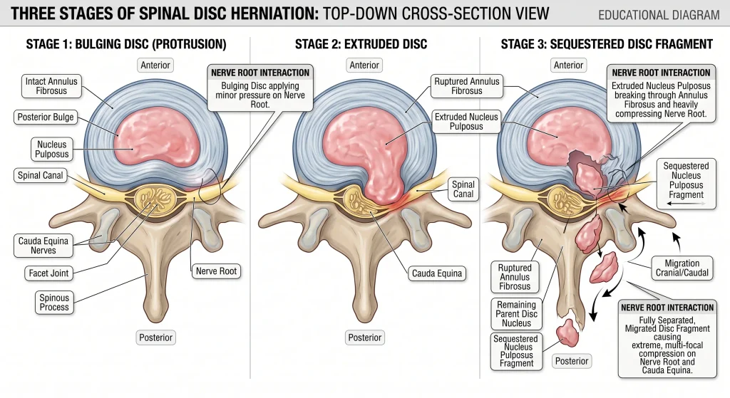
A systematic review published in Clinical Rehabilitation pooled imaging studies of people managed conservatively and found that spontaneous regression occurred in 96% of sequestrated discs and 70% of extruded discs, compared with 41% of protrusions and 13% of bulges. Complete resolution was reported in 43% of sequestrations and 15% of extrusions. Those are not marginal numbers, and they describe people who did not have surgery.
What is actually happening inside the disc when it herniates?
A spinal disc is a pressurised joint, not a cushion. It has a tough outer wall of concentric collagen rings called the annulus fibrosus, and a hydrated gel core called the nucleus pulposus. A herniation happens when the core pushes through a fissure in those rings. The displaced material can then press on a nearby nerve root, chemically irritate it, or both — which is why symptoms rarely stay confined to the back.
Those collagen rings are laid down at alternating angles, which makes them very good at resisting compression and rotation and considerably less good at resisting repeated bending under load. That is why the classic mechanism is not a single heroic lift but a hundred unremarkable ones: bending to load a dishwasher, lifting a car seat out of a back seat, a long drive followed immediately by unloading a boot. Disc pressure rises with sustained flexion, and a wall that has been quietly fissuring for years can give way during something entirely ordinary.
The symptom pattern usually follows the mechanics. Pain that intensifies with sitting, bending forward and getting out of a car, and eases with walking or lying flat, is a fairly typical mechanical disc pattern. A sudden electric spike through the leg with a cough, a sneeze or a hard strain is one of the more characteristic signs, because those actions momentarily raise pressure around the nerve root. Where nerve root involvement is present, the leg symptoms often carry a specific quality — burning, electric, or a dead-feeling heaviness — and the particular sensations people describe often say more about which structure is involved than the pain score does.
Why do larger herniations often heal better than small bulges?
Because resorption depends on exposure. A sequestered fragment — one that has separated completely from the disc and sits free in the epidural space — is fully surrounded by vascular tissue and immune cells, so the breakdown process can work on all of it. A contained bulge is still inside the annulus, shielded from that response. The frightening word on the report often describes the herniation most likely to disappear.
This single fact reframes an enormous number of scan reports. Patients routinely arrive convinced that “extrusion” is a worse diagnosis than “bulge,” and on the imaging it certainly looks more dramatic. Biologically, the opposite ranking often applies. The 96% regression figure for sequestration against 13% for bulging is not a rounding error — it reflects a real mechanistic difference in how much material the immune system can actually reach.
It also explains a clinical pattern that confuses people: someone with a small bulge and a stubborn, grumbling back that never fully settles, and someone else with an alarming extrusion whose leg pain resolves in four months. The larger herniation had more inflammatory material to clear, which hurt more at the start — and more of it was accessible, which is why it cleared.
What does “healed” actually mean for a disc?
Healing here means the herniated material has been cleared and the nerve is no longer irritated — not that the disc has been restored to its original condition. The tear in the annulus repairs with disorganised fibrous scar tissue rather than regenerating the original layered architecture, and disc height that was lost generally stays lost. The realistic target is a back that tolerates load without symptoms, not a scan that reads clean.
This distinction matters more than it sounds, because it decides what you measure success against. If the yardstick is a normal MRI, many people who have made a full functional recovery will feel they have failed. Scar tissue in the annulus is mechanically decent but not identical to the original — slightly stiffer, less elastic, and it is why some people notice a residual sensitivity to deep end-range bending for a long time after the leg pain has gone.
The useful yardsticks are functional: how long you can sit before symptoms escalate, how far the symptoms travel down the leg, how much you can lift, and whether an ordinary day costs you the following morning. Those move well before an MRI changes, and they are what actually determines quality of life.
How long does it take, and what should you notice along the way?
Recovery is not one process on one timeline. Chemical irritation around the nerve usually settles first, over weeks. Physical resorption of the herniated material takes longer, often three to twelve months. Annular repair runs on its own schedule again, and sensory and motor recovery are slower still. Knowing which process you are waiting on tells you whether slow progress is normal or worth investigating.
| What is changing | Typical timeframe | What you notice | What it means if it stalls |
|---|---|---|---|
| Chemical inflammation settling around the nerve root | 2 to 6 weeks | Leg pain stops being constant and starts responding to position changes | Pain that is unchanged at 6 weeks with no position that eases it warrants reassessment |
| Centralisation — symptoms retreating up the limb | 2 to 8 weeks | Foot and calf symptoms fade before the buttock and back do | Symptoms travelling further down the leg rather than up is the wrong direction and should be assessed |
| Physical resorption of the herniated material | 6 weeks to 12 months, most change at 3 to 6 months | Steadily longer sitting, standing, driving and walking tolerance | Tolerance flat for 6 or more weeks despite genuine graded loading suggests a review of the plan |
| Annular defect closing with fibrous scar | 3 to 6 months | Coughing, sneezing and bending stop producing sharp spikes | Repeated sharp flares from trivial load at 6 months suggests ongoing instability of the segment |
| Sensory recovery — numbness, pins and needles | 3 months to 2 years, sometimes incomplete | The numb patch shrinks and its edges become vague rather than sharply defined | No change at 12 months often means a permanent patch, which is usually a nuisance rather than a disability |
| Motor recovery — weakness, foot drop | 3 to 12 months, least predictable | Return of calf raises, heel walking and confident push-off on stairs | No measurable strength gain by around 3 months is a reason to seek a surgical opinion, not to keep waiting |
Two honest caveats. First, these ranges are wide because the biology genuinely varies — fragment size, how much of it is exposed to vascular tissue, and individual inflammatory response all shift the curve. Second, symptom improvement and imaging change do not run in step. Many people feel substantially better long before the scan changes, and some scans improve while a sensitised nervous system keeps symptoms going. Neither situation means the process has failed.
Why do numbness and weakness lag behind the pain?
Pain is largely driven by inflammation and pressure, both of which resolve relatively quickly. Numbness and weakness reflect actual damage to nerve fibres, and nerve tissue repairs slowly — regenerating axons advance at roughly a millimetre a day, which for a lumbar nerve supplying the foot means months. Longstanding or severe compression can leave a residual deficit even after the disc material has completely cleared.
In practice this produces a sequence that surprises people: the leg pain fades, the person feels dramatically better, and then they notice the numb patch on the outside of the calf or the top of the foot is still exactly where it was. That is normal and not a sign the recovery has stalled. Sensory fibres are thinner and more vulnerable, so they tend to be affected earlier and recover later.
Weakness deserves closer attention. A mild, improving weakness that tracks alongside pain relief usually reflects pain inhibition rather than nerve damage, and it recovers quickly once loading resumes. Weakness that is measurable — you cannot do a single-leg calf raise, you cannot heel-walk, your foot slaps on the stairs — and is not improving over the first six to twelve weeks is a different conversation, and the timing of that conversation genuinely influences the outcome.
When should you stop waiting and get assessed?
Waiting is appropriate when symptoms are mechanical, position-responsive and trending in the right direction. It is not appropriate when there are signs that the nerve supply itself is being compromised, or when the picture does not behave like a disc at all. The features below each point to something that does not follow the usual resorption timeline, and each needs prompt assessment rather than patience.
- Difficulty passing urine, loss of bladder or bowel control, numbness around the inner thighs, genitals or buttocks, or new sexual dysfunction. This combination may indicate cauda equina syndrome — compression of the bundle of nerve roots below the spinal cord. It is time-critical and requires same-day emergency assessment, not a physical therapy appointment.
- Weakness that is worsening, or that affects both legs. Progressive or bilateral motor loss suggests compression that is increasing rather than settling, which changes the clinical urgency entirely.
- Foot drop, or an inability to push off or lift the front of the foot. A defined motor deficit needs formal assessment early, because the length of time a nerve has been compressed is one of the factors that influences how completely it recovers.

- Fever, unexplained weight loss, a history of cancer, immunosuppression, injected drug use, or a recent spinal procedure. These raise the possibility of infection or malignancy, which do not follow disc rules and do not resorb.
- Severe pain following significant trauma, or sudden onset in someone with osteoporosis or long-term steroid use. Vertebral fracture can present with similar pain but requires a completely different management pathway.
- Pain that is unrelenting at rest and unaffected by any position, particularly at night. Mechanical disc pain almost always has a position of relief. Pain that ignores position is a signal to look beyond the disc.
Quick check
Five yes-or-no questions. Any “yes” in the first two means seek emergency care today.
- Has your ability to control your bladder or bowel changed at all since this started?
- Is there any numbness in the area that would contact a bicycle saddle?
- Can you rise onto the ball of one foot ten times on the affected side without your heel dropping?
- Over the past two weeks, have the symptoms moved further down the leg rather than further up?
- Is there any position at all — lying, standing, walking — that reliably reduces the pain?
Does an MRI tell you whether your disc will reabsorb?
Partially. An MRI can show the type of herniation, and type does predict resorption likelihood — extrusions and sequestrations regress far more often than contained bulges. What a scan cannot do is tell you how much pain you will have, how fast you will recover, or whether you need surgery. Imaging describes structure; it does not measure symptoms, and the two correlate far more loosely than most people assume.
That gap is the reason imaging is not the default first step. National guidance from the National Institute for Health and Care Excellence advises against routinely offering imaging for low back pain and sciatica outside a specialist setting, and recommends it only where the result would change what happens next. Disc changes, degeneration and even herniations show up regularly on scans of people with no symptoms whatsoever, so a finding on its own does not establish the source of pain.
Imaging genuinely changes management in a narrow set of situations: when red flags are present, when a motor deficit is progressing, when the symptom distribution does not match the suspected level, or when surgery or a nerve root injection is being actively considered and the surgeon needs to confirm that the compression matches the symptoms. Outside those, a scan mostly adds vocabulary — which is why learning what the phrases in an imaging report actually mean clinically is often more useful than the scan itself.
What actually helps this along, and what does nothing?
Resorption is an immune process, and no treatment currently available has been shown to speed it up directly. What management can do is control the symptoms while it happens, protect the nerve from repeated provocation, and keep the surrounding system strong enough that you return to full function once the material clears. Movement, sensible load management and adequate sleep support that. Passive treatment alone does not.
What has genuine support: continuing normal activities as far as symptoms allow, rather than resting in bed; graded exercise, particularly as things move from the acute phase into the recovery phase; and position management so that the nerve is not provoked for hours at a time. Sleep deserves a specific mention, because sleep deprivation measurably lowers pain thresholds — a disc flare that wrecks your sleep creates a feedback loop where poor sleep amplifies the pain that is causing the poor sleep.
What has not been shown to accelerate resorption: inversion tables, home spinal decompression units, collagen or “disc regeneration” supplements, and repeated passive treatment on its own. Some of these feel good in the moment, and short-term symptomatic relief has value. The distinction worth holding onto is between relief and repair. Hands-on treatment is best used as part of a package that includes exercise rather than as a standalone plan, which is the practical difference between the various passive treatment options people compare and a program that actually changes load tolerance.
One myth worth dismantling directly: the idea that a herniated disc can “slip back into place” if it is manipulated, hung from, or stretched correctly. Displaced nuclear material does not return through a torn annulus the way a dislocated joint relocates. It is cleared, not repositioned. Any treatment sold on the promise of pushing it back in is describing a mechanism that does not exist.
From the Clinic: Dr. Arora’s Expert Insight
Week five is where I lose people. Not week one — week one is frightening enough that nobody argues about doing the exercises. Week five is different. The leg pain has faded to a background hum, the person decides they are basically fine, goes back to a full day at a desk or lifts something entirely unremarkable, and wakes the next morning with the calf symptom back. That flare arrives at exactly the moment their confidence had started to rebuild, and the conclusion they draw is that the whole thing has failed. A meaningful proportion of the people who stop rehabilitation stop in that week, on the back of a recovery that was working.
Here is what I try to get across before that week arrives. A flare at week five is almost never resorption running backwards — the immune process does not reverse because you sat too long. It is a load-tolerance mismatch: capacity was improving, and the demand jumped past it faster. The thing I ask people to record is not a pain score but the furthest point down the leg the symptoms reach, checked once a day in one line. After a flare, that number is what tells you whether the trajectory has changed. If the symptoms spiked hard but stayed above the knee when they used to reach the foot, the recovery is intact and you have simply found the current ceiling. Pain intensity after a flare is noisy. Distribution is not.
The other thing that reliably goes wrong is the advice to “strengthen your core,” delivered without any thought about which direction the disc failed in. People come in having done weeks of crunches and sit-ups, which are repeated loaded lumbar flexion — the precise mechanical direction that provoked the herniation in the first place. They have been diligently rehearsing the injury mechanism and wondering why the leg symptoms will not settle. What works better in this specific condition is building trunk control in positions that do not repeatedly flex the lumbar spine under load — anti-rotation and anti-extension work, loaded carries, and a hip hinge trained until it is genuinely automatic — so that the strength you gain is strength in the pattern that failed, not strength in the pattern that broke it.
What should you do in the first 72 hours?
The goal in the first few days is not to fix anything — it is to stop provoking the nerve while the inflammatory phase peaks. That means avoiding long stretches in any single position, especially sustained sitting, keeping movement frequent but short, and finding two or three positions that reliably reduce symptoms so you can rotate between them. Complete bed rest is not the answer, but neither is pushing through.
What to do today
- Change position every 20 to 30 minutes, before the symptoms build — not after.
- Walk for 5 to 10 minutes several times a day rather than once for an hour.
- Find your relief position and use it deliberately — often lying with the knees supported, or standing and gently extending. Test both; use whichever moves symptoms up the leg.
- Brace before you sneeze or cough: stand up if you can, put a hand on a surface, and tighten the abdomen first. This is the cheapest flare prevention available.
- Use heat or ice based on which one actually helps you — for symptom control only, in 15-minute blocks.
- Protect sleep aggressively. A side-lying position with a pillow between the knees keeps the lumbar spine neutral overnight.
- Do not repeatedly test the painful movement to see if it still hurts. Each test re-provokes the nerve, and checking every hour is one of the most common self-inflicted setbacks.
How do you rebuild from there?
Progression should be driven by what your symptoms do over the following 24 hours, not by the calendar. The general sequence is: calm the nerve, restore basic tolerance to sitting, standing and walking, retrain the hip hinge so bending stops loading the lumbar spine, then add load in that pattern. Advancing before a stage is stable is the single most common cause of the week-five setback.
| Phase | Main goal | What it looks like | Ready to progress when |
|---|---|---|---|
| 1 — roughly weeks 0 to 2 | Reduce nerve provocation without going still | Short frequent walks, position rotation, gentle preference-direction movement, no sustained flexed sitting | Leg pain is no longer constant and you can sit around 20 minutes without a symptom spike |
| 2 — roughly weeks 2 to 6 | Rebuild everyday tolerance and hinge mechanics | Walking 20 to 30 minutes, hip hinge with a dowel, glute bridges, side plank progressions, dead bugs | A 30-minute walk causes no next-day flare, and you can hinge to mid-shin without leg symptoms |
| 3 — roughly weeks 6 to 12 | Load the pattern that failed | Goblet squats, Romanian deadlifts from blocks, farmer carries, split squats, resisted anti-rotation work | You can carry roughly half your bodyweight for 30 metres (about 100 feet) with symptoms staying below buttock level the next day |
| 4 — 3 months onward | Return to sport, heavy lifting or physical work | Progressive deadlift and squat loading, running return, rehearsing the specific lifts your job or sport demands | Full-range loaded hinge without symptoms, 20 or more single-leg calf raises on each side, and two consecutive weeks without symptom escalation |
A few condition-specific rules are worth stating plainly. Do train the hip hinge before you train anything heavy — if your hips do not share the bending load, the lumbar segment takes it. Do break up driving on long journeys, since a reclined seat plus vibration plus sustained flexion is one of the more reliable ways to provoke an irritable root. Do keep a structured, repeatable set of movements rather than improvising daily, which is where a consistent home exercise program earns its place.

Equally: do not do crunches, sit-ups or loaded toe-touches in the first three months. Do not stretch into the position that reproduces the leg symptoms in the belief that you are freeing something. Do not sit for an hour to “prove” you can. And do not chase a repeat MRI to confirm resorption when your function is clearly improving — the scan will not change what you do next, and a report that still shows a herniation in someone who feels well causes more anxiety than it resolves.
For return to activity, use functional milestones rather than dates. Driving: you can perform an emergency stop without hesitation or a symptom spike. Desk work: 40 minutes of sitting without escalation, with breaks planned rather than reactive. Gym: a full-range unloaded hinge with no leg symptoms before any bar is added. Running: 30 minutes of brisk walking plus 20 single-leg calf raises per side, then a short jog-walk trial, with the following morning as the real verdict.
What if it does not settle?
A meaningful minority of herniations do not follow the expected curve. If leg pain and function have not improved after roughly six to twelve weeks of genuine, well-executed conservative care — not six weeks of rest and hoping — that is the point to escalate rather than repeat the same plan for another three months. Escalation usually means a specialist assessment, a targeted nerve root injection, or a surgical opinion.
Surgery for a disc herniation is an operation on the nerve’s environment, not a repair of the disc. Decompression removes the fragment that is compressing the root, which is why it works well when the compression is the problem and works poorly when it is not. National guidance frames this clearly: spinal decompression is considered for people whose pain or function has not improved with non-surgical treatment and whose imaging findings correspond to their symptoms. Both conditions matter — a scan finding that does not match the clinical picture is not a surgical target.
The two situations where the decision moves faster are a progressive motor deficit and suspected cauda equina syndrome. Outside those, choosing surgery is usually a decision about how much longer you are willing to wait for a process that would probably complete on its own — a legitimate trade-off, and one worth making deliberately rather than in a panic during a bad week.
The realistic outlook
So, can a herniated disc heal on its own? For most people, yes — and the more dramatic the herniation looks on the report, the better the odds that the body will clear it. Resorption is a real, well-described biological process, and it does the heavy lifting while good management keeps you functional and prevents you from repeatedly re-irritating the nerve while it works.
The practical step, starting today: pick one measure and track it daily in a single line — the furthest point down your leg that symptoms reach. Not a pain score. If that point is retreating up the limb across two to three weeks, your recovery is on track and the plan does not need changing. If it is descending, or if any of the red flags above appear, book the assessment rather than waiting out another month.
Frequently asked questions
Can a herniated disc come back after it heals?
Re-herniation at the same level is possible, but it is not the usual outcome. The repaired annulus is scar tissue rather than original tissue, so that segment can stay somewhat more sensitive to heavy, repeated end-range bending. Building genuine hinge mechanics and hip strength is the most useful protection available.
Will my MRI ever look completely normal again?
Often not entirely, and that is not a problem. Herniated material can resorb fully, but reduced disc height, signs of degeneration and the scarred annular defect commonly remain visible. Plenty of people with permanently abnormal-looking scans have no symptoms at all.
Is it safe to keep lifting weights with a herniated disc?
Usually yes, with the load and the movement direction adjusted rather than the gym abandoned. Pressing, carrying and hip-dominant work can often continue much earlier than people expect, while loaded spinal flexion and maximal deadlifting should wait. The test is what your symptoms do the following morning, not during the set.
If numbness never fully goes away, does that mean permanent damage?
A residual patch of altered sensation can persist and does represent some permanent change to sensory fibres, but it rarely affects function. What matters clinically is strength and balance, not whether a coin-sized area of the foot feels slightly different. Numbness that is spreading, rather than static, is the version that needs review.
Do I need a follow-up MRI to confirm the disc has reabsorbed?
Not if you are getting better. Repeat imaging is worthwhile when symptoms are not improving, when a neurological deficit is progressing, or when an intervention is being planned. Scanning purely for reassurance frequently backfires, because degenerative findings that were always there get read as new problems.
Can a herniated disc heal without any physical therapy at all?
The resorption itself is immune-driven and happens regardless of whether you attend appointments. What rehabilitation changes is the time spent in pain, how much strength and tolerance you lose along the way, and how likely you are to keep re-provoking the nerve while the fragment clears. The disc may resolve either way; the recovery experience differs considerably.
If I avoid surgery now, will I just need it later anyway?
Choosing conservative care first does not commit you to surgery later, and it does not make later surgery harder. The main exception is a progressive motor deficit, where waiting can affect how completely the nerve recovers. Absent that, a trial of conservative management is a reasonable and reversible first choice.
Medical disclaimer
This article explains how herniated disc material is cleared by the body and what recovery generally looks like. It cannot tell you whether your particular symptoms are coming from a disc, which spinal level is involved, or whether the timeline described here applies to you — those require a hands-on neurological examination that no article can replace. Nothing here should be used to delay assessment of bladder or bowel changes, saddle numbness, or leg weakness that is getting worse. If you are already under the care of a clinician for this problem, their assessment of your specific presentation takes precedence over any general guidance in this article.
References
- Chiu CC, Chuang TY, Chang KH, Wu CH, Lin PW, Hsu WY. The probability of spontaneous regression of lumbar herniated disc: a systematic review. Clinical Rehabilitation. 2015;29(2):184-195. doi:10.1177/0269215514540919
- National Institute for Health and Care Excellence. Low back pain and sciatica in over 16s: assessment and management (NG59). Published 2016, last updated 2020.