You are holding a report with words like “narrowing,” “canal compromise” or “compression” in it, and the appointment to discuss it is two weeks away. That gap is where most of the worry happens. So here is the part nobody tells you first: the finding on the scan is not the diagnosis. What decides whether it matters is what your legs do when you walk.
Spinal stenosis on MRI means the spaces inside your spine — the central canal, the lateral recesses, or the openings where nerves exit — have narrowed. It is extremely common with age and is frequently found in people with no symptoms at all. The finding becomes clinically meaningful when it matches a specific pattern: leg heaviness or cramping that builds with walking and eases within minutes of sitting or leaning forward.
Key takeaways
- Narrowing on a scan is not the same as a problem. Degenerative spinal findings are present in a high proportion of people with no back pain, and become more common with every decade.
- The symptom pattern is what counts. Neurogenic claudication — leg symptoms provoked by walking and relieved by forward flexion — is the clinical picture stenosis actually produces.
- Flexion relief is a clue, not a lifestyle. Leaning forward eases symptoms, but spending all day slumped weakens the hips and trunk and shrinks your walking tolerance further.
- Conservative care is the default first step. Most people improve their function substantially without surgery, and the narrowing itself does not need to change for that to happen.
- Some signs do change the timeline. Progressive weakness, foot drop, or any bladder or bowel change moves this from a rehabilitation problem to an urgent one.
What does spinal stenosis on MRI actually mean?
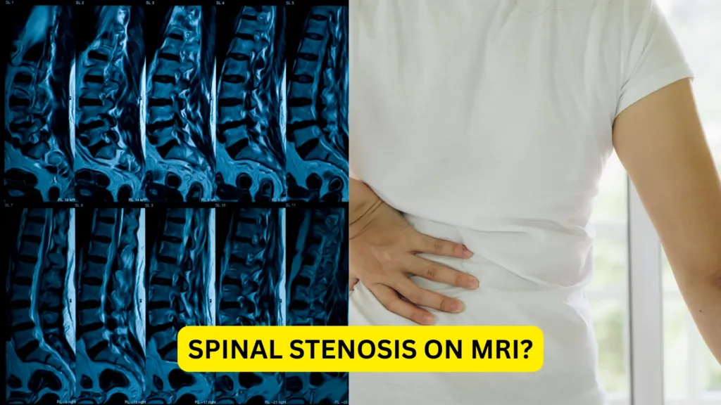
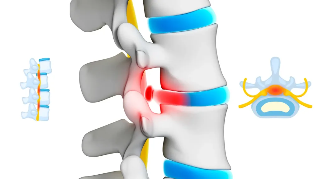
It means the space available for nerve tissue inside your spine has been reduced — usually by a combination of a thickened ligament, an enlarged facet joint, a bulging disc, and a disc that has lost height. Radiologists grade it mild, moderate or severe based on how much room is left. That grade describes anatomy. It does not describe how you feel or how you will do.
This distinction is the single most useful thing to understand about your report. A systematic review of imaging in people with no back symptoms, published by Brinjikji and colleagues in the American Journal of Neuroradiology, found disc degeneration in 37% of pain-free 20-year-olds rising to 96% by age 80, and disc bulges in 30% rising to 84% over the same span. Their conclusion was blunt: many of these findings are part of normal aging and are not associated with pain.
Stenosis follows the same logic. Narrowed canals are found routinely in people walking around without complaint. A useful way to think about it: the scan tells you how wide the road is, not how much traffic is stuck on it.
Types of Spinal Stenosis
Lumbar Spinal Stenosis
This is the most common type and affects the lower back. It can cause:
- Pain radiating down the legs (sciatica-like symptoms)
- Tingling or numbness in feet or calves
- Leg fatigue or heaviness when walking or standing
- Relief when leaning forward or sitting down (shopping cart sign)
It often limits walking distance and can mimic peripheral vascular disease, so a thorough diagnosis is essential.
Cervical Spinal Stenosis
This affects the neck region and can be more serious. Why?
Because your spinal cord passes through the cervical spine. If it’s compressed, symptoms can involve:
- Neck pain with radiating arm symptoms
- Loss of hand dexterity
- Balance issues
- In severe cases, weakness or paralysis
This type of stenosis deserves prompt evaluation and management.
Causes of Spinal Stenosis
A Spinal stenosis MRI typically reveals one or more of the following culprits:
- Aging: Natural wear-and-tear leads to degeneration of discs, joints, and ligaments
- Herniated Discs: Discs that bulge or rupture may intrude into the spinal canal
- Bone Spurs (Osteophytes): Extra bone growths can reduce space in the canal
- Thickened Ligaments: Ligaments like the ligamentum flavum may stiffen and thicken
- Spinal Injuries: Fractures or trauma can shift bone or cause swelling
- Congenital Conditions: Some people are born with a naturally narrower canal
Understanding the root cause helps guide your treatment and prognosis.
Risk Factors You Should Know
You’re more likely to be diagnosed with spinal stenosis on MRI if:
- You’re over 50 years old
- Have osteoarthritis or rheumatoid arthritis
- Lead a sedentary lifestyle
- Are overweight
- Have had prior spinal surgery or injuries
That said, younger individuals—especially athletes and those with heavy lifting jobs—are not immune, especially if structural changes exist from previous injuries or congenital narrowing.
What does the wording on my report mean?
Radiology reports describe location and degree, not consequence. A phrase names which structure has narrowed, at which spinal level, and by roughly how much. None of it tells you whether that narrowing is producing your symptoms — that comes from the examination. Here is what the common phrases are actually saying.
- “Central canal narrowing at L4-L5”: The main tunnel for your spinal cord is tight at the L4-L5 vertebral level (lower back).
- “Mild foraminal stenosis on the right”: Nerve passageways (foramina) are narrowed, possibly pinching nerve roots.
- “Disc bulge with facet hypertrophy”: The disc is protruding, and the small joints in the spine are enlarged from degeneration.
These terms help determine the severity and location of your stenosis, which in turn influences treatment planning.
Common Terms in MRI Reports
- Degenerative Disc Disease: Age-related disc changes, often seen by age 30-40.
- Disc Bulge: A disc is pushing out but not ruptured.
- Facet Joint Arthropathy: Arthritis of spine joints causing pain and stiffness.
- Foraminal Narrowing: Openings where nerves exit are reduced in size.
The words “severe” and “marked” are the ones that frighten people most, and they describe a measurement rather than your prognosis. Plenty of people with severe narrowing on paper walk normally, and some with mild narrowing have genuinely limiting symptoms. The report has to be read alongside what you can and cannot do.
Is the stenosis actually causing my symptoms?
Compare your pattern against neurogenic claudication, which is what symptomatic lumbar stenosis produces. Leg heaviness, cramping or burning builds as you walk, worsens when you stand upright, and settles within a few minutes of sitting or leaning forward on a cart or counter. If your pain does not behave that way, the narrowing on your report may be incidental — and treating it as the cause will waste months.
| Feature | Neurogenic claudication (stenosis) | Vascular claudication (arterial) | Mechanical low back pain |
|---|---|---|---|
| What brings it on | Walking, and standing still while upright | Walking a fairly consistent distance; standing still is fine | Bending, lifting, sustained sitting, specific movements |
| What relieves it | Sitting, or leaning forward — the position matters more than stopping | Simply stopping; position does not matter | Position change, unloading, sometimes walking |
| Cycling uphill or pushing a cart | Usually comfortable, because the trunk is flexed forward | Provokes symptoms as much as walking does | Variable |
| Stairs | Going up often easier than going down — climbing flexes the spine | Going up harder, because demand on the legs rises | Both may aggravate depending on the driver |
| Symptom quality | Heaviness, cramping, pins and needles, often both legs but asymmetric | Tight, cramping calf pain, reliably in the same muscle group | Ache or sharp catch, usually back-dominant |
| Feet and pulses | Normal pulses; skin and nails normal | Weak or absent foot pulses, cool skin, hair loss on the legs | Normal |
If your legs behave like the middle column, the problem is arterial rather than spinal, and no amount of spine rehabilitation will help. That single distinction is worth making early.
Symptoms You May Be Feeling
Early-Stage Signs
- Aching in lower back or neck
- Occasional tingling in arms or legs
- Subtle weakness when climbing stairs or gripping objects
Progressed Symptoms
- Cramping in calves or thighs after walking (neurogenic claudication)
- Pain that improves when bending forward or sitting
- Gait imbalance or frequent stumbling
Signs that need urgent assessment
Stenosis usually progresses slowly, which is why sudden change is the thing to watch. Each of the following suggests that nerve tissue is under pressure severe enough that time to treatment affects the outcome. Do not wait for a scheduled appointment.
- New loss of bladder or bowel control, difficulty starting urination, or numbness across the groin and inner thighs. This pattern suggests compression of the nerve bundle at the base of the spinal cord. Recovery of bladder function depends heavily on how quickly pressure is relieved, which is why this is a same-day emergency rather than an urgent referral.
- Rapidly progressing weakness in a leg or foot — a foot that slaps the floor, or catching a toe on steps. Weakness that is getting measurably worse over days indicates ongoing nerve damage rather than nerve irritation, and the window for full recovery narrows as it continues.
- New clumsiness in the hands, dropping objects, or a change in balance and walking. In cervical stenosis the spinal cord itself is involved, and cord compression produces these signs before pain. This warrants prompt neurological assessment even when the neck barely hurts.
- Numbness that is spreading in area rather than staying in one strip. An expanding sensory loss suggests the level of compression is increasing, which changes the urgency of imaging and review.
- Severe symptoms after a fall or collision, particularly with osteoporosis or a cancer history. Fracture and other structural causes need excluding before symptoms are attributed to degenerative narrowing.
Does a stenosis finding mean I need surgery?
No. Conservative care is the standard first step for lumbar spinal stenosis, and most people never progress past it. Surgery becomes a serious consideration when a well-executed rehabilitation program has failed to improve walking tolerance, when neurological deficits are developing, or when daily function has narrowed to a point you are not prepared to live with.
The sequence generally runs: activity and load modification, targeted physical therapy, medication where useful, then injections, and only then a decompression procedure. The National Institute of Neurological Disorders and Stroke maintains a plain-language overview of the condition if you want an independent reference point before your appointment.
Who Should You See First If You See Spinal Stenosis On MRI: Neurologist, Orthopedist, or PT?
Here’s a smart sequence:
- Physiatrist (non-surgical spine specialist)
- Physical Therapist (physiotherapist) (for customized rehab plan)
- Orthopedic or Neurosurgeon (if symptoms progress or fail to improve)
The key is team-based care. One-size-fits-all rarely works with spinal issues.
Getting a Second Opinion
Always wise, especially when:
- Surgery is suggested early
- You’re unsure about diagnosis
- Symptoms don’t align with MRI findings
A second (or third) set of eyes ensures you’re not missing safer, effective options.
Non-Surgical Management Options
Lifestyle Modifications
- Posture correction: Use lumbar support, adjust screen height
- Movement breaks: Avoid prolonged sitting or standing
- Weight management: Reduces spinal load
- Sleep hygiene: Use supportive pillows; back sleeping with knee elevation is ideal
Small daily changes make a big long-term difference.
Physical Therapy and Exercise
Physical therapy (physiotherapy) for stenosis is not generic back rehabilitation. The whole program is built around one biomechanical fact: extending the lumbar spine closes the canal further, and flexing it opens the canal up. Exercise selection follows from that.
- Flexion-biased mobility. Single and double knee-to-chest, seated forward lean, and posterior pelvic tilts all increase canal dimension temporarily and often reduce leg symptoms within a minute or two.
- Hip extensor and hip flexor work. Tight hip flexors pull the pelvis into anterior tilt, which extends the lumbar spine and narrows the canal all day long. Strong glutes take load off the spine during walking, which is precisely the activity that provokes symptoms.
- Trunk endurance in a neutral or slightly flexed position. Endurance matters more than maximal strength here, because symptoms appear during sustained upright activity rather than during a single lift.
- Interval walking, deliberately structured. Walk until leg symptoms begin, take a 30–60 second forward-lean or sit break, then continue. Total daily distance rises far faster this way than by attempting one long uninterrupted walk.
- Cardio that keeps the spine flexed. Stationary cycling, walking on an incline (which naturally leans you forward), and pool walking are usually well tolerated when flat overground walking is not.
What consistently aggravates stenosis is sustained or repeated lumbar extension — standing back bends, prone press-ups, overhead lifting with an arched low back, and long periods of standing still. Note that this is the opposite of what helps disc-related pain, which is one reason a program borrowed from a friend with a disc problem so often makes stenosis worse.
Progression is judged by walking tolerance, not by pain scores. If the distance you can cover before leg symptoms start is increasing week to week, the plan is working — even if your back still aches at the end of the day.
👨⚕️ Dr. Arora’s Clinical Note:
A common mistake I see is turning the “shopping cart sign” into a full-day posture strategy—because the short-term relief is so convincing. Clinically, that advice is incomplete. Yes—gentle flexion often reduces leg symptoms (the classic “shopping cart” relief), but the mistake is living in a constant slumped position all day. That can decondition the trunk and hips, stiffen the thoracic spine, and make walking tolerance worse over time.A more reliable approach is the “Flexion Sweet Spot + Capacity Build” rule:
- Use flexion as a symptom tool, not a posture. Do short resets (30–60 seconds) of supported forward-lean or a brief sit break only when symptoms start.
- Then build capacity in the hips and trunk (glutes + core endurance) so your spine doesn’t take the full load. Often, improving hip extension strength reduces the “back doing everything” pattern that fuels stenosis flare-ups.
- Try this quick self-check: Walk 3 minutes upright, note symptoms; then walk 3 minutes with a slight forward lean (hands on thighs or a treadmill rail). If the lean gives clear relief, you likely benefit from interval walking (walk until symptoms begin → 30–60 sec lean/reset → repeat) rather than long continuous walks that flare you.
If you notice new or worsening leg weakness, foot drop, numbness that spreads quickly, or bowel/bladder changes, stop self-management and seek urgent medical evaluation.
Pain Management with Medications
Common pharmacological options:
- NSAIDs: Ibuprofen, naproxen—for inflammation
- Muscle relaxants: For spasms
- Neuropathic pain agents: Gabapentin or pregabalin target nerve pain
These are not long-term fixes but can ease symptoms during rehab phases.
Epidural Steroid Injections – Pros & Cons
Pros:
- Often provide fast relief (especially leg symptoms)
- Allow participation in physical therapy
Cons:
- Temporary (effects last weeks to months)
- Risk of complications with repeated use
- Not effective for all patients
Discuss timing and frequency with a pain management specialist.
When Surgery Becomes Necessary
Surgery is typically considered when:
- Conservative care fails
- Neurological deficits develop (e.g., weakness, foot drop)
- Daily life is significantly limited
Surgical Options Available
- Laminectomy: Removes part of the vertebra (lamina) to relieve pressure
- Discectomy: Removes herniated disc material
- Spinal Fusion: Stabilizes vertebrae by joining them together
Each has pros and cons depending on your age, level of stenosis, and health.
Recovery Time and Expectations
- Minimally invasive procedures: Resume light activity in 2–6 weeks
- Fusion surgeries: May need 3–6 months of supervised rehab
Physical therapy post-op ensures you regain strength and mobility safely.
Minimally Invasive Surgeries
These newer techniques offer:
- Shorter hospital stays
- Smaller incisions
- Less blood loss
- Faster recovery
Always ask your surgeon if you’re a candidate.
Living With Spinal Stenosis
Daily Tips for Comfort
- Use supportive seating with lumbar rolls
- Take standing/walking breaks every 30–60 minutes
- Heat therapy: Warm baths, hot packs can ease muscle tension
What to be cautious with
- Prone extension work — press-ups, superman holds, back extension machines. These close the canal further and commonly increase leg symptoms in stenosis, even though they help many other back conditions.
- Overhead pressing with an arched low back. The arch, not the weight, is the problem. The same lift is usually fine with the ribs down and the pelvis neutral.
- Long periods of standing still. Standing at a counter or in a line provokes symptoms more reliably than walking does, because there is no relief phase.
- Heavy loading before hip strength is rebuilt. Deadlifts and loaded squats are not permanently off the table, but they belong later, once walking tolerance and hip extensor strength have improved.
What to do this week
1. Measure your baseline: walk at a comfortable pace and note the time or distance at which leg symptoms first appear. Write it down.
2. Replace one long walk with three or four short interval walks, each stopping just before symptoms start.
3. Add two minutes of knee-to-chest or seated forward lean whenever leg symptoms build, rather than pushing through.
4. Start hip extensor work — bridges and sit-to-stands — three days this week.
5. Re-measure your baseline in seven days. A rising number is the outcome that matters, not the wording on your report.
Long-Term Outlook
The good news? Most people with spinal stenosis live full, active lives.
Key takeaways:
- Don’t delay early treatment
- Build spinal strength and flexibility
- Monitor changes and consult professionals
Consistency wins over quick fixes.
The bottom line
Spinal stenosis on MRI describes a narrowing that a very large number of people carry without difficulty. Whether it matters in your case is decided by how your legs behave when you walk, how far you can go before symptoms start, and whether leaning forward changes anything. Those three observations tell a clinician more than the adjective in your report.
If your pattern matches neurogenic claudication, the work is specific and it usually pays off: open the canal with position, build the hips so the spine carries less, and expand walking tolerance in intervals rather than in one heroic effort. If your pattern does not match, the more useful next step is finding out what does — before anyone operates on a finding that was never the problem.
FAQs
1. Is spinal stenosis on MRI curable without surgery?
Not curable, but absolutely manageable with therapy, posture correction, and medication.
2. How long can you live with spinal stenosis?
A full, healthy life—most people don’t need surgery.
3. What worsens spinal stenosis?
Poor posture, inactivity, weight gain, and ignoring early symptoms.
4. Can spinal stenosis go away on its own?
The narrowing stays, but symptoms can improve or stabilize.
5. What sleeping position is best for spinal stenosis?
Back sleeping with knees slightly elevated or side sleeping with a pillow between the knees.
6. Is spinal stenosis a form of arthritis?
It often results from arthritis but is not itself arthritis.
7. Is walking good for spinal stenosis?
Yes! Especially on flat surfaces with short, frequent intervals.
8. What’s the difference between spinal stenosis and a herniated disc?
A herniated disc is one cause of spinal stenosis—stenosis is the result of narrowed spaces.
9. Are epidural injections safe for spinal stenosis?
Generally safe when done by specialists, but effects may be temporary.
10. Can physical therapy really help spinal stenosis?
Yes, especially when personalized and consistent—it’s a cornerstone of non-surgical care.
References
- Brinjikji W, Luetmer PH, Comstock B, et al. Systematic literature review of imaging features of spinal degeneration in asymptomatic populations. American Journal of Neuroradiology. 2015;36(4):811–816. doi:10.3174/ajnr.A4173 (PMID: 25430861)
- Munakomi S, Cruz R. Lumbar Spinal Stenosis. In: StatPearls [Internet]. Treasure Island (FL): StatPearls Publishing; 2024. NCBI Bookshelf NBK531493
With 20+ years of clinical experience in spine, joint, and pain management, Dr. Vivek ensures every article reflects accurate, trustworthy, and up-to-date health advice.
Learn more about Dr. Vivek’s qualifications & journey →