Expert Review: This article includes clinical insights from Dr. Vivek Arora, a physical therapist (physiotherapist) with 20+ years of experience.
A disc problem at this level rarely announces itself politely. It often starts as a deep, dull ache across the low back after a weekend of bending, lifting, or sitting badly in a car seat. Then, a few days later, a line of pain travels down one leg and finishes somewhere unexpected — the outside of the calf, the top of the foot, or the big toe. L4-L5 disc herniation symptoms follow a reasonably predictable map, because the map is drawn by which nerve root is being irritated and from which direction. Understanding that map will not give you a diagnosis, but it will tell you what your body is trying to report, which parts of it deserve attention today, and which parts can safely be given time.
Key takeaways
- Direction matters more than size. A herniation pushing backward and slightly to one side usually irritates the L5 nerve root; one pushing far out to the side can catch the L4 root instead.
- The L5 pattern ends at the big toe. Buttock, outer thigh, outer calf, across the top of the foot, into the great toe — with weakness lifting the foot and the big toe.
- The L4 pattern stays in front. Front and inner thigh, across the kneecap, down the inner shin — with weakness straightening the knee and a reduced knee reflex.
- Progressive foot drop is not a wait-and-see symptom. Weakness that is measurably worse week on week warrants prompt assessment rather than another month of patience.
- Most cases settle without surgery. Leg pain from a lumbar disc commonly improves over weeks to a few months, and imaging findings alone rarely change that path.
What is an L4-L5 disc herniation, and why does this level fail so often?
An L4-L5 disc herniation happens when the soft inner core of the disc sitting between the fourth and fifth lumbar vertebrae pushes through a weakened outer ring and presses on or chemically irritates a nearby nerve root. This level is one of the two most commonly involved in the lower back because it sits where a mobile lumbar spine begins handing load down toward a relatively fixed pelvis, absorbing high bending and shearing forces in the process.
A lumbar disc is not a cushion in the pillow sense. It behaves more like a sealed, water-filled bag under pressure. The centre, called the nucleus pulposus, is a gel that redistributes load in whatever direction the spine is being bent. The outer wall, the annulus fibrosus, is a ring of tough fibres laid down in criss-crossing layers, and it is those layers that fail. Repeated forward bending under load pushes the nucleus backward against the rear wall of the annulus. Add rotation, and only about half the annular fibres are oriented to resist that twist at any given moment, which is why the classic story is bending and turning at the same time rather than lifting something heroically heavy in a straight line.
The L4-L5 segment carries a specific burden. Below it, L5-S1 is anchored by thick iliolumbar ligaments and the sacrum, which is essentially bolted into the pelvis. That relative stiffness means L4-L5 becomes the most freely moving segment for flexion in many people, and it also absorbs a large share of shear — the forward-sliding force that builds when the trunk tips over the hips. Pressure inside the disc rises further with sitting, with forward bending, and momentarily with anything that spikes abdominal pressure, which is why some people first notice the problem as a sudden jolt of back pain during a cough or sneeze rather than during a lift.
This pattern also explains who tends to present with it. Herniation at L4-L5 shows up most often in adults roughly between their thirties and mid-fifties, when the nucleus still holds enough water to migrate under pressure but the annulus has accumulated enough wear to give way. In older adults, the same disc has usually dried and flattened, and leg symptoms are more likely to come from narrowing of the nerve channels than from a fresh herniation.
Which nerve root does an L4-L5 herniation actually press on?
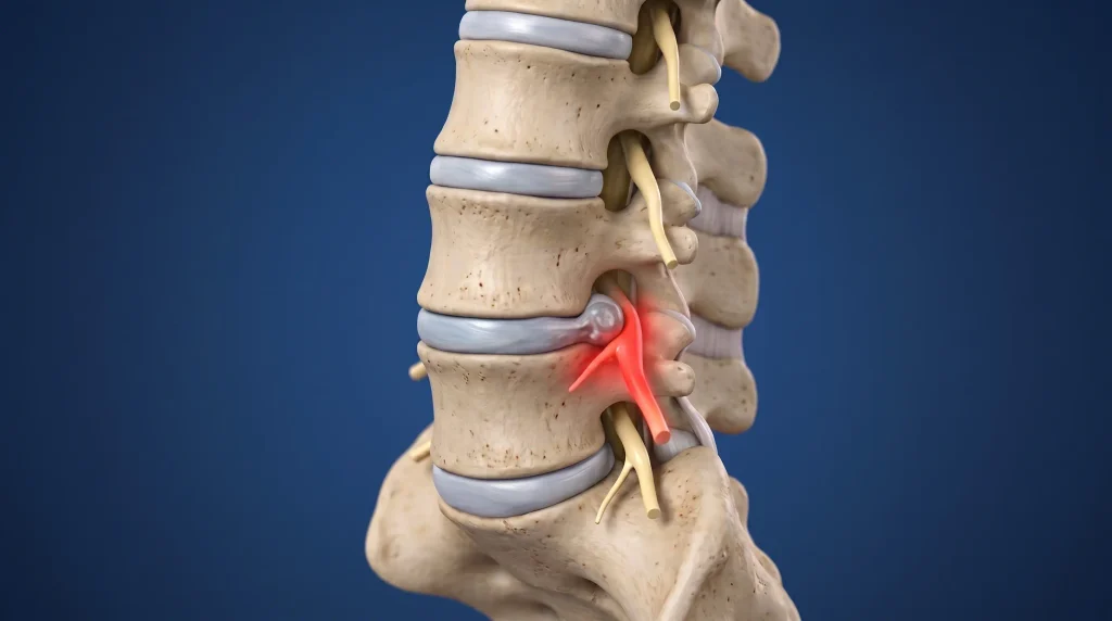
It depends on the direction the disc material travels. Most L4-L5 herniations move backward and slightly to one side, where they meet the L5 nerve root passing down toward its exit one level below — so an L4-L5 herniation usually produces L5 symptoms, not L4 ones. A less common far-lateral herniation, escaping sideways beyond the spinal canal, catches the L4 root as it leaves the spine and produces a completely different symptom map.
The reason this trips people up is anatomical. At every lumbar level, two sets of nerve roots are in play. The exiting root — at L4-L5 that is L4 — is already curving outward toward its doorway high in the side wall of the segment. The traversing root — L5 — is still travelling downward through the canal, hugging the back corner of the disc, on its way to leave one level lower. A herniation aimed at that back corner meets L5 first. It has to travel much further out to the side, into or beyond the doorway itself, before it reaches L4.
- Posterolateral or paracentral herniation — the common one. Compresses the traversing L5 root. Expect the buttock-to-big-toe pattern.
- Far-lateral or foraminal herniation — uncommon. Compresses the exiting L4 root. Expect front-of-thigh and inner-shin pain, and often more pain on standing and extending than on sitting.
- Large central herniation — can involve several roots at once and, if it is big enough to crowd the whole bundle of nerves in the canal, raises the concern of cauda equina compression, which is a medical emergency.
L4-L5 disc herniation symptoms: what you feel and where
Nerve root irritation produces three separable things: pain along a strip of skin, altered sensation such as numbness or pins and needles in that same strip, and weakness in the specific muscles that root supplies. The L5 pattern runs down the outside of the leg and finishes on the top of the foot and the big toe. The L4 pattern runs down the front and inner side of the thigh, crosses the knee, and finishes on the inner shin.
| Nerve root | Where you feel it | What tends to get weak | What to check at home | What a positive check may suggest |
|---|---|---|---|---|
| L5 (usual root affected at L4-L5) | Buttock, outer thigh, outer calf, across the top of the foot, into the big toe | Lifting the foot upward (ankle dorsiflexion), lifting the big toe, pushing the hip out sideways | Walk 10 steps on your heels with the toes lifted; try to hold your big toe up while pressing down on it with your thumb; stand on one leg in front of a mirror for 30 seconds | Heel walking collapses on one side, the big toe gives way easily, or the opposite hip visibly drops — suggests L5 motor involvement |
| L4 (far-lateral herniation) | Front and inner thigh, across the kneecap, down the inner shin toward the inner ankle | Straightening the knee (quadriceps), controlling the knee on descent | Stand up from a low chair on one leg without pushing with your arms; walk down four stairs leading with that leg | The knee buckles, wobbles, or drops the last few degrees — suggests L4 motor involvement. The knee reflex needs a clinician to test |
| S1 (shown for contrast — usually an L5-S1 problem) | Buttock, back of thigh, back of calf, heel, sole, little toe | Pushing off with the toes (calf), single-leg balance during push-off | Perform 15 to 20 single-leg calf raises on each side and compare | Fewer repetitions or a lower heel height on one side points away from L4-L5 and toward the level below |
Now the honest caveat, because this is where symptom maps get oversold. Dermatomes — the strips of skin each nerve root supplies — vary considerably between individuals and overlap heavily at their borders. Published maps disagree with each other, and a real person’s pain rarely respects a clean line drawn on a diagram. Pain that spills across the boundary between the L5 and S1 territories does not mean you have two herniations, and pain that fizzles out at the ankle instead of reaching the toe does not rule an L5 root problem in or out. Weakness in a specific muscle group is generally a more reliable signal than the exact geography of the ache. The particular qualities of nerve-related leg pain — the electric, burning, or cord-like character of it — are also more informative than where the line happens to stop.
What else can produce this pattern?
Several conditions borrow parts of the L4-L5 script, and telling them apart changes what you do next.
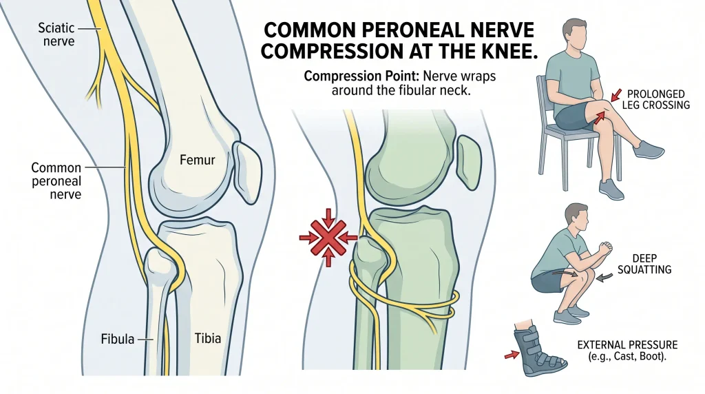
- Peroneal nerve compression at the knee. The common peroneal nerve wraps around the outside of the fibula just below the knee and is easily squashed by prolonged leg crossing, deep squatting, or a tight cast or brace. It causes foot drop and numbness on the top of the foot, which looks exactly like L5. The separating features: it usually comes with no back pain at all, hip abduction strength stays normal, and turning the foot inward is typically preserved because that muscle is supplied through a different branch.
- Deep gluteal or piriformis-type irritation. Buttock-dominant pain that worsens with prolonged sitting on a hard surface and eases with standing can mimic proximal L5 symptoms, but it rarely produces true muscle weakness or a crisp toe-level pattern. The distinction between buttock-driven pain and genuine nerve root compression is worth making early, because the treatment emphasis differs.
- Hip joint pathology. Hip osteoarthritis classically refers pain to the groin and front of the thigh, overlapping the L4 map. Hip pain usually moves with hip rotation and worsens with weight bearing; L4 root pain tends to track with spinal positions and often includes numbness on the inner shin, which a hip never causes.
- Lateral recess or foraminal narrowing. In adults past their fifties, the same root can be pinched by thickened ligament and bone rather than by disc material. The giveaway is often a pattern that builds with walking and standing and eases within a minute or two of sitting or leaning forward.
Why does the back pain sometimes dominate before the leg pain shows up?
Because the disc wall itself has a nerve supply, and it can hurt long before anything touches a nerve root. The outer third of the annulus carries small nerve fibres, so a tear in that wall can produce severe, deep, central low back pain on its own. Only when disc material migrates far enough to contact or chemically inflame the root does the leg pattern appear — which may be days or weeks later, or never.
This sequence explains a change that alarms a lot of people. In the first phase, back pain is the loud symptom and the leg is quiet. Then the back pain settles somewhat and the leg pain flares. It feels like deterioration, and sometimes it is — but it can also simply be the shift from a wall-pain problem to a root-irritation problem as the herniation reaches its final position.
The direction of travel is the useful thing to watch, and it goes the other way too. When leg symptoms retreat up toward the buttock and back over days or weeks, even if the back itself feels temporarily worse, that is generally a favourable trend. When symptoms march the other way — from the buttock down toward the calf and foot, or from ache into numbness and weakness — that deserves attention. Keep a simple daily note of the furthest point down the leg that you felt anything. It is cruder than any test, and far more informative than a pain score out of ten.
Which symptoms should not be watched and waited on?
Most leg pain from an L4-L5 herniation is safe to manage patiently. A small number of features are not. The level-specific one here is progressive weakness of the foot and big toe — a foot drop that is measurably worse this week than last. The others are the classic signals of nerve bundle compression, fracture, infection, or disease elsewhere. Each earns its place for a specific reason.
- Worsening foot drop. A foot drop is difficulty lifting the front of the foot, so the toes catch on kerbs, stairs, and carpet edges. A stable, mild weakness that is not changing can often be managed conservatively alongside the pain. Weakness that is clearly deepening over days to weeks suggests ongoing compression, and the longer a motor nerve is under pressure the less predictable its recovery becomes. This one should be assessed promptly rather than given another month.
- Numbness in the saddle area, difficulty passing or controlling urine, loss of bowel control, or new sexual numbness. These suggest compression of the cauda equina, the bundle of nerve roots below the end of the spinal cord. This is a surgical emergency and warrants same-day emergency assessment, not an appointment next week.
- Symptoms in both legs at once, particularly if they appeared quickly. A single herniation usually affects one side. Bilateral involvement raises the possibility of a large central herniation crowding the canal.
- Fever, chills, night sweats, or unexplained weight loss with back pain. These point away from a mechanical disc problem and toward infection or another systemic cause that needs different investigation entirely.
- Severe pain after a fall or impact, especially with osteoporosis or long-term steroid use. This raises the question of fracture, which changes the immediate management and the imaging decision.
- A personal history of cancer with new, unrelenting back pain that does not vary with position. Mechanical pain almost always has positions that help. Pain with no easing position at all is worth investigating.
Seek care if
- You could heel walk ten steps last week and cannot manage five this week.
- Your toes have started catching on stairs, kerbs, or the edge of a rug.
- Numbness is spreading rather than staying in the same patch.
- Leg pain has been steadily peripheralising — travelling further down the limb — for more than two weeks.
- Any saddle numbness or bladder or bowel change appears. This one is same-day emergency care.
When is an MRI actually needed for an L4-L5 herniation?
An MRI is worth doing when the result would change what happens next — most often when someone has persistent radicular leg pain despite a reasonable course of conservative treatment and is being considered for an injection or surgery, or when a red flag suggests something other than a straightforward disc. In the first several weeks of typical leg pain with no alarming features, a scan usually confirms what the examination already showed and does not alter the plan.
UK guidance from the National Institute for Health and Care Excellence advises against routine imaging for low back pain and sciatica in non-specialist settings, and recommends imaging only in specialist care where the result is likely to change management. That is not a cost-cutting position. It reflects a genuine clinical problem: disc bulges, protrusions, and annular changes at L4-L5 appear on scans of people with no pain at all, and they become more common with age. A scan that finds one in a person who happens to have back pain has not necessarily found the cause.
What imaging does add, when it is indicated, is precision about things the examination cannot see: whether the herniation is paracentral or far-lateral, whether it is contained within the annulus or has broken through, whether a fragment has migrated up or down behind the vertebral body, and whether the nerve channels are additionally narrowed by degenerative change. Those details matter if a targeted injection or a decompression is being planned. They matter far less if the plan is graded rehabilitation. This is also why reading a report cold can be so unsettling — the vocabulary of narrowing and degenerative change on a lumbar MRI sounds far more ominous than the clinical picture usually is.
Three things people get wrong about L4-L5 herniations
Some of the most persistent beliefs about lumbar disc herniation are not just unhelpful — they actively steer people toward worse decisions. Three come up more than the rest: that the size of the herniation predicts the outcome, that a foot drop means an operation is inevitable, and that rest is the treatment. None of them holds up well against how these cases actually behave.
Myth one: a bigger herniation means a worse outcome. The relationship between herniation size and symptom severity is weak. A small fragment sitting directly against an inflamed root can be agonising, while a large one that has migrated into space where there is room to spare can be nearly silent. Larger extruded fragments also tend to reduce in size over time, since the immune system treats displaced disc material as something to clear away.
Myth two: any foot drop means surgery. Mild, stable dorsiflexion weakness alongside improving pain is frequently managed conservatively with good results. It is the trajectory that drives the decision, not the presence of weakness in itself. Dense, rapidly worsening weakness is a different conversation, and a time-sensitive one.
Myth three: rest until it goes away. Here the evidence deserves an honest reading rather than a slogan. A Cochrane review comparing advice to rest in bed with advice to stay active found that for acute low back pain, staying active gave small benefits in pain and function — but for sciatica specifically, it found little or no difference between the two. So the case for keeping moving with an irritated L5 root is not that movement dissolves the herniation. It is that prolonged bed rest costs you conditioning, sleep quality, confidence, and time, while giving nothing measurable back. NICE guidance likewise encourages people to continue with normal activities as far as possible.
From the Clinic: Dr. Arora’s Expert Insight
The most useful sentence in an L4-L5 assessment is usually the one said sideways — dropped casually while someone is sliding off the plinth and reaching for their shoes, long after the formal history is finished. A version of the same offhand remark surfaces again and again: that one shoe is scuffing at the toe far faster than the other, or that a sandal keeps flicking off on that side, or that the treadmill belt catches the front of that foot now. None of it is offered as a symptom. It is offered as small talk about footwear. And it is frequently the first observable evidence of dorsiflexor weakness, arriving weeks before anyone thinks to test heel walking.
That matters because of how generic disc advice is structured. The standard package for a lumbar herniation is built almost entirely around pain: avoid bending, manage flare-ups, wait it out, come back if the pain worsens. Pain is the variable everyone tracks. But at L4-L5, the clinically decisive variable is often motor, and motor decline is quiet. It does not hurt more. It sometimes hurts less, because a root under sustained pressure can stop generating pain while it is still losing conduction. I have seen the pain-only script produce a genuinely reassured patient whose foot was steadily getting weaker, and the reassurance was the problem — nobody had been asked to check strength, so nobody noticed. The other failure mode I see is the reflexive extension-based programme handed to every disc patient. For the common backward-and-to-the-side herniation it often helps. For a far-lateral one narrowing the L4 doorway, repeated extension can close down the very space the root needs, and the person dutifully doing thirty of them a day gets steadily worse while following instructions correctly.
What works better is embarrassingly simple. I ask people to run a thirty-second motor check on the same day each week and write the result on their phone: how many steps they can heel walk before the foot flops, whether the big toe can resist a firm thumb, and whether the pelvis stays level standing on that leg for half a minute. Three numbers, once a week. It converts a vague sense of things being worse into a trend line that tells us whether patience is justified or whether the situation has changed. And I test directional preference rather than assume it — I want to see what repeated bending backward and forward actually does to the leg symptoms over a few minutes before anyone is given a programme built on one of them.
What should you do in the first 72 hours?
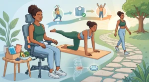
The goal in the first few days is not to fix the disc — it is to calm nerve irritation, avoid the positions that keep provoking it, and stay in enough motion to protect your conditioning and sleep. That means short and frequent movement rather than either bed rest or pushing through. It also means establishing a baseline for the strength checks that will tell you, over the coming weeks, whether things are moving in the right direction.
What to do today
- Record your baseline. Heel walk steps, big toe resistance, single-leg pelvis level. Write down the numbers and the date.
- Break up sitting every 20 to 30 minutes. Disc pressure is high in unsupported sitting, and the L4-L5 segment carries the most of it. A one-minute stand and walk is enough.
- Walk in short bouts. Five to ten minutes, several times a day, rather than one long attempt. Stop when leg symptoms travel further down, not when the back merely aches.
- Find two positions of ease. Lying on your back with knees supported on pillows, or side-lying with a pillow between the knees, will suit most people. Use them for genuine rest, not for the whole day.
- Protect the first hour after waking. Discs absorb fluid overnight and are at their most pressurised on waking. Delay deep bending, shoe-tying from standing, and loaded lifting until you have been up and moving for 30 to 60 minutes.
- Discuss pain relief with a clinician rather than self-escalating. Nerve-related pain responds differently from muscular pain, and the choice of medication is not interchangeable.
How does rehab progress from acute pain to loaded movement?
Rehabilitation for an L4-L5 herniation advances on criteria, not on the calendar. Each phase has a job: settle root irritation, then rebuild tolerance to sitting, standing, and walking, then restore strength in the specific muscles the affected root supplies, then reintroduce hinging and loading. You move forward when your symptoms behave a certain way for several consecutive days — not because a given number of weeks has passed.
| Phase | Main goal | What it looks like in practice | Ready to progress when |
|---|---|---|---|
| 1. Settle (roughly the first 1 to 2 weeks) | Reduce root irritation and reclaim basic daily function | Frequent short walks; positions of ease; testing which direction of repeated movement draws symptoms out of the leg; gentle nerve gliding within a pain-free range | Leg symptoms no longer travel past the knee at rest, and a 10-minute walk does not leave you worse for the rest of the day |
| 2. Tolerate (roughly weeks 2 to 6) | Build sitting, standing and walking tolerance; begin targeted strength | Hip hinge patterning with a dowel; isometric trunk work with a neutral spine; side-lying hip abduction and side planks for the L5-supplied gluteus medius; heel walks and tibialis raises for dorsiflexion | You can sit 30 minutes or walk 20 minutes without leg symptoms increasing, and heel walking is roughly symmetrical for 20 steps |
| 3. Load (roughly weeks 6 to 12) | Restore strength under load through the hips and trunk | Goblet squats, split squats, controlled step-downs, farmer carries around 10 to 20 kg (22 to 44 lb), progressive hip hinge with light load | No next-morning symptom flare after a loaded session, single-leg stance holds the pelvis level for 30 seconds, and big toe strength matches the other side |
| 4. Return (3 months onward) | Rebuild capacity for sport, heavy work, and unplanned load | Graded return to deadlifting, rowing, running, or occupational lifting; speed and fatigue reintroduced last | You can complete a full session at target load with symptoms unchanged 24 hours later, across two or three consecutive sessions |
The exercise selection is not arbitrary. Hip abduction work earns its place because the gluteus medius is largely L5-supplied, and a hip that drops during single-leg stance is both a sign of that weakness and a source of ongoing rotational stress at the very segment you are trying to settle. Dorsiflexion loading — heel walks, tibialis raises — targets the muscles most likely to be affected and gives you a strength number you can track. Hinge patterning matters because the mechanism that provoked the herniation was almost certainly a spine that bent when the hips should have. A structured programme of physical therapy exercises for the lower back is worth more than any single movement in isolation, and the sequence matters as much as the selection.
Do’s and don’ts specific to this level
- Do track dorsiflexion strength weekly. It is the single most decision-relevant number in an L4-L5 problem.

- Do get the driver’s seat right if you commute. Sustained sitting with the pelvis rolled backward is one of the highest-pressure positions for this disc, and a small lumbar support plus a slightly reclined backrest changes it meaningfully.
- Do use the direction of symptom travel as your progress measure rather than pain intensity. Leg symptoms retreating toward the buttock is progress even if the back feels sorer for a few days.
- Don’t do repeated end-range flexion in the first hour after waking — toe-touch stretches, sit-ups, or loading the dishwasher first thing.
- Don’t assume repeated extension is automatically correct. It helps many backward-and-to-the-side herniations and can aggravate a far-lateral one. Judge it by what your leg symptoms do over the following few minutes and hours.
- Don’t keep hard-stretching a hamstring that feels tight on the symptomatic side. That tightness is frequently protective nerve tension rather than short muscle, and aggressive stretching pulls on an already irritated root.
- Don’t resume loaded hinging — deadlifts, heavy rows, lifting children from the floor — until single-leg pelvic control and toe strength are symmetrical.
Return-to-activity milestones
Useful milestones are things you can pass or fail, not feelings. Desk work generally becomes sustainable once you can sit 30 minutes with a standing break and no leg symptom increase. Driving beyond about 30 minutes is reasonable when you can perform an emergency-stop movement without hesitation and the affected foot has full, confident dorsiflexion. Running is a phase-four activity: symmetrical heel walking, 20 single-leg calf raises each side, and pain-free walking of 45 minutes come first. Loaded lifting returns last, at roughly half your previous working weight, progressing only if the following morning is unchanged.
What happens when conservative care stalls, and when does surgery come in?
Surgery becomes a genuine conversation when leg pain and functional limitation persist despite an adequate course of non-surgical care, and when the imaging findings clearly correspond to the symptoms and examination. NICE recommends considering spinal decompression for sciatica when non-surgical treatment has not improved pain or function and radiological findings are consistent with the symptoms. It is a decision about matching, not about the presence of a herniation on a scan.
Before that point, a stall is worth diagnosing rather than simply waiting out. Genuine plateaus usually have a reason: the programme has been built around the wrong direction of movement; sitting load at work has never actually been reduced despite good intentions; sleep has been broken for weeks, which reliably amplifies pain sensitivity; or the strength deficits were never targeted because everything focused on pain. Reassessment at six to eight weeks of no change should look at all four before it concludes that conservative care has failed.
The urgency picture is different from the elective one. Cauda equina features require emergency assessment. A rapidly progressing motor deficit — a foot drop deepening over days — is a prompt-referral situation rather than a wait-and-review one, because the relationship between how long a motor root is compressed and how completely it recovers is not a favourable one. By contrast, pain alone, however severe, is rarely an emergency, and severity of pain is a poor guide to whether surgery is needed.
As for outlook: the majority of people with radicular leg pain from a lumbar disc improve substantially over weeks to a few months without an operation, and displaced disc material often reduces in size over that period. A discectomy — removing the fragment pressing on the root — tends to relieve leg pain faster than waiting does, but the difference between the two paths narrows considerably over a year or two. That is precisely why the decision usually comes down to how much the leg pain is costing you now, and whether the deficit is stable, rather than to what the scan shows.
The bottom line on L4-L5 disc herniation symptoms
The pattern of L4-L5 disc herniation symptoms is a map of which root is under pressure and from which direction — usually L5, giving you the outer-leg-to-big-toe line with weakness lifting the foot; occasionally L4, giving you the front-of-thigh and inner-shin line with a weaker knee. That map is a guide, not a diagnosis, because dermatomes overlap and real pain ignores diagrams.
Here is the practical instruction to leave with. Today, record three numbers: how many steps you can heel walk before the foot flops, whether your big toe can hold against firm thumb pressure, and whether your pelvis stays level standing on the affected leg for thirty seconds. Repeat them the same day every week. Improving or stable numbers mean patience and graded loading are doing their job. Numbers going the wrong way mean it is time to be assessed, regardless of what your pain is doing.
Frequently asked questions
Can you have an L4-L5 disc herniation with no leg pain at all?
Yes, and it is common. A herniation that has not reached or inflamed a nerve root may produce only deep central back pain, stiffness, and difficulty bending, or no symptoms whatsoever — herniations are found routinely on scans of people with no complaints. Leg pain appears only when the root is mechanically compressed or chemically irritated.
How long does foot drop from an L4-L5 herniation take to recover?
Recovery of a mild, stable foot drop commonly takes several weeks to several months once the compression settles, because nerve fibres regenerate slowly. The strongest predictors are how severe the weakness was at its worst and how long the root stayed compressed, which is why a deepening foot drop is treated as time-sensitive rather than something to monitor indefinitely.
Is an L4-L5 herniation worse than an L5-S1 herniation?
Neither level is inherently worse — they simply produce different symptom maps and different functional problems. An L4-L5 herniation more often affects the L5 root, so weakness shows up in lifting the foot and stabilising the hip. An L5-S1 herniation more often affects S1, so it shows up in calf push-off and single-leg spring.
Can I keep training at the gym with an L4-L5 disc herniation?
Usually yes, with the exercise menu changed rather than emptied. Machine-supported and upright work, carries, and single-leg strength are typically tolerable early; heavy spinal flexion under load is not. Returning to barbell lifting after a lumbar disc herniation works best as a staged rebuild rather than a return to previous weights.
Why does my leg pain feel worse when I sit but better when I walk?
Sitting raises pressure inside the lumbar disc and rolls the pelvis backward, which increases the load on the rear wall of the disc where most L4-L5 herniations sit. Walking reduces that pressure and moves the nerve through its sheath. A sitting-worse, walking-better pattern is fairly typical of disc-related root irritation.
Does a herniated disc at L4-L5 heal, or do I have it permanently?
The disc does not return to its original architecture, but the displaced material often shrinks over months as the body breaks it down, and symptoms usually settle well before any structural change is visible. Most people end up functionally normal with a disc that still looks abnormal on imaging, which is a far better outcome than it sounds.
Can an L4-L5 herniation cause groin or front-of-thigh pain?
It can, if the herniation is far-lateral and irritates the exiting L4 root, which supplies the front and inner thigh. Because hip joint problems refer pain to the same area, the distinguishing features are numbness on the inner shin and knee-extension weakness, which point to the nerve root rather than the hip.
Medical disclaimer
The symptom maps, home strength checks, and progression criteria described here are educational tools for understanding what a lumbar nerve root problem may look like — they are not a diagnosis, and they cannot distinguish an L4-L5 herniation from the several other conditions capable of producing similar leg symptoms. Nerve root involvement needs a hands-on neurological examination, and the decisions that matter most in this condition, particularly those around progressive weakness, imaging, injections, or surgery, require an individual assessment by a qualified clinician who has examined you. If you develop numbness in the saddle area, any change in bladder or bowel control, or rapidly worsening weakness in the foot or leg, seek emergency medical care immediately rather than acting on anything written here.
References
- National Institute for Health and Care Excellence. Low back pain and sciatica in over 16s: assessment and management (NG59). Published 2016, last updated 2020. https://www.nice.org.uk/guidance/ng59
- Dahm KT, Brurberg KG, Jamtvedt G, Hagen KB. Advice to rest in bed versus advice to stay active for acute low-back pain and sciatica. Cochrane Database of Systematic Reviews. 2010;(6):CD007612. https://doi.org/10.1002/14651858.CD007612.pub2