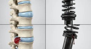
Expert Review: This article includes clinical insights from Dr. Vivek Arora, a physical therapist (physiotherapist) with 20+ years of experience.
A lumbar disc herniation is one of the few spine diagnoses that sounds like it should end in an operation and usually does not. Recovering from a herniated disc without surgery is the normal route, not the consolation prize — but it works far better run as a staged plan than left as a vague instruction to rest and hope. A herniation means the tough outer ring of a spinal disc has torn enough to let some of the softer inner material push outward, where it can compress or chemically irritate a nearby nerve root. What follows is the whole journey in four stages, each advancing on a functional criterion rather than a date.
Key takeaways
- Most disc-related sciatica settles without an operation. Herniated material is gradually reabsorbed, so time plus well-directed loading beats time alone.
- Progress by criteria, not dates. Advance when you can do a specific thing without leg symptoms worsening.
- Centralization is the signal to watch. Symptoms retreating from calf toward buttock means the plan is working, even if the back feels more sore.
- Imaging rarely changes the early plan. A scan earns its place when the result would alter management.
- Stopping rehab the week the pain stops is the commonest relapse trigger. Pain resolves weeks before load tolerance returns.
What is actually happening when a disc herniates?
A spinal disc is a pressurized cushion between two vertebrae: a firm layered outer ring called the annulus, holding a gel-like core called the nucleus. A herniation occurs when those layers fail in one spot and some of the core migrates outward. Pain then comes from two mechanisms at once — mechanical pressure on a nerve root, and an inflammatory chemical reaction around it. That chemical component is why symptoms often fail to track bulge size, and why the first fortnight can improve before anything structural has changed.
Biomechanically, the annulus handles compression well and sustained loaded flexion poorly. Sitting slumped drives nuclear material backward, toward exactly where a nerve root exits — which is why long drives and repeated bending over a low sink flare things, while walking and lying ease them.
Why do most herniated discs improve without an operation?
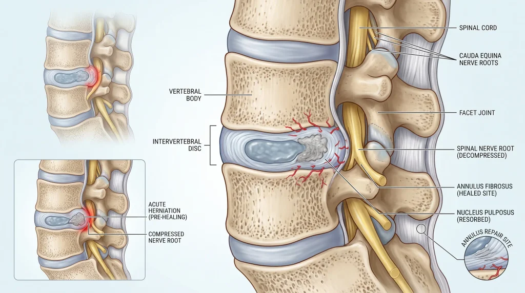
This image is for educational purposes only and does not substitute for professional medical advice, and may not be accurate.
The body treats herniated disc material as displaced tissue. Over weeks to months the immune system breaks it down and reabsorbs it, inflammation subsides, and the nerve root regains space. Conservative care exists to protect that process, keep you moving, and rebuild capacity so the same tissue is not overloaded again. Discs herniate most often in the thirties, forties and fifties, while the nucleus is still hydrated enough to migrate.
How do I know whether my leg pain is really from a disc?
Disc-related nerve root pain travels in a defined band down the leg, often past the knee, and is provoked by sitting, bending, coughing or sneezing. It usually comes with pins and needles or numbness in a strip of skin, and eases with walking or lying down. Pain that stays in the buttock, shifts with hip position, or hurts on direct pressure over the glutes points somewhere else.
Quality matters as much as location: radicular pain is electric or burning and follows a line rather than a region — the sensations that identify sciatica rather than a muscular ache. Two look-alikes recur. Deep gluteal irritation bothers the sciatic nerve along its course rather than at the root, and separating piriformis-type symptoms from true nerve root sciatica changes what belongs in your program. Hip pathology is the other, producing groin and lateral thigh pain people mislabel as back pain.
Which symptoms mean you should not wait?
Most disc symptoms are unpleasant rather than dangerous. A small number are not. Each feature below suggests either something other than a settling nerve root, or a root under enough pressure that time is no longer neutral. None confirms a serious problem alone, but each shifts the decision from watchful management to prompt assessment.
- Numbness around the genitals or inner thighs, or difficulty starting or controlling urination. This raises the possibility of cauda equina compression, where the nerve bundle at the base of the spinal cord is squeezed — an emergency, because the window for preserving bladder and sexual function is short.
- Weakness that is worsening rather than stable. A foot catching on stairs means motor fibers are affected. Stable mild weakness is often monitored; progressive weakness is not, because the compression is winning.
- Symptoms in both legs at once. One herniation usually irritates one root, so bilateral symptoms suggest central compression or a non-disc cause.
- Fever, night sweats, or back pain after a recent infection or procedure. Spinal infection is rare, but it produces pain that does not obey mechanical rules and needs blood tests rather than exercise.
- A history of cancer, unexplained weight loss, or pain unrelieved by any position. Mechanical disc pain almost always has a position that helps; pain with none deserves investigation.
- Significant trauma, or a minor strain with osteoporosis or long-term steroid use. These raise the possibility of vertebral fracture, which changes early management entirely.
Seek care today if
- You cannot feel the toilet seat properly, or cannot tell when your bladder is full.
- Your foot has become noticeably weaker over the past few days.
- Leg symptoms have spread from one leg to both.
- Pain is severe and unrelieved in every position, including lying down.
The first item is an emergency department problem, not a wait-and-see problem.
Do you actually need an MRI, and does a repeat scan help?
For most people in the early weeks, no. Imaging is worth doing when the result would change what happens next — when serious pathology needs excluding, or an injection or operation is genuinely being considered. National guidance on low back pain and sciatica advises against routine imaging in non-specialist settings, reserving it for situations where the result is likely to alter management.
An MRI confirms which level is involved and whether compression matches the symptom distribution — decisive for a surgeon, largely irrelevant in week two of a settling flare. Scans also pick up incidental findings in people who feel entirely well, so a report listing bulges and annular tears at several levels does not automatically explain your pain. Repeat scanning is where time and money quietly disappear: a second MRI almost never changes the plan in an improving patient. The exceptions are new or progressing neurological loss, a genuinely changed symptom pattern, or a surgical decision needing current anatomy.
Which beliefs about disc injuries slow recovery down?
Four do the most damage: that a herniated disc means a permanently damaged spine, that bending must be avoided forever, that treatment can push the disc back into place, and that pain must reach zero before strengthening can begin. None hold up. The disc is living tissue that remodels, bending is a movement the spine is built for once tolerance is rebuilt, no manual technique repositions nuclear material, and rehabilitation that waits for silence never starts. The related trap is treating passive care as the treatment itself — hands-on work and manipulation can make movement accessible, but they stop being useful the moment they become the entire plan.
From the Clinic: Dr. Arora’s Expert Insight
If I were allowed only one piece of information about a new patient with a herniated disc — the MRI report, or their honest answer to what they expect to be doing in three months — I would take the answer, every time. It never appears on a scan, but what someone believes is going to happen shapes how they move, how much they hold back, and whether they turn up for week four. A Cochrane prognostic review found that in non-specific low back pain, individual recovery expectations are probably strongly associated with future work participation — a broader population than disc herniation, but it matches what walks through the door.
Here is the specific way generic advice fails this condition. People are told to avoid bending and to strengthen the core, and they follow both faithfully for months. The result is a rigid, braced trunk on hips that have quietly lost the ability to hinge. The first time they reach into a car boot or lift a child from a cot, all the movement has to come from the lumbar spine, because it is the only segment still willing to move. Bracing without hip contribution does not protect a disc — it concentrates load on it. What gets missed early is not the disc at all; it is how much bend a person can produce at the hip before the lumbar spine takes over.
A pattern that comes up repeatedly is the person who improves quickly, feels good around week five, and stops. They are not being careless — the pain has genuinely gone. But pain resolution and load tolerance recover on different timelines, and the second lags by weeks. When they come back it is almost never a new injury; it is the same disc, re-provoked by an ordinary task their capacity had not caught up to.
The four-stage plan for treating a herniated disc without surgery
Recovery runs in four stages: settle the irritation, restore movement and unloaded strength, rebuild capacity under load, then return to full demand and hold it. The week ranges are typical, not prescriptive — progression is decided by the exit criterion in the right-hand column, and a stage taking twice as long as its range is common rather than a failure.
| Stage | Main goal | Core work | Ready to progress when |
|---|---|---|---|
| 1. Settle the irritation (weeks 0–2) | Reduce nerve root irritation, restore daily function | Frequent short walks, directional preference, sitting and sleep management | Leg symptoms sit above the knee, you sit 20 minutes and walk 10 minutes without a spike, and sleep through most of the night |
| 2. Movement and unloaded strength (weeks 2–6) | Recover hip and trunk control, desensitize the nerve | Nerve gliding, glute and hip work, dead bug and side plank, unloaded hinge | Symptoms confined to back and buttock, 30 minutes continuous walking, and a hinge to mid-shin without symptoms travelling further down the leg |
| 3. Rebuild capacity (weeks 6–12) | Restore load tolerance in the patterns real life demands | Loaded hip hinge, squat pattern, carries, graded rehearsal of work tasks | You lift from the floor at roughly half bodyweight without leg symptoms, carry a moderate load 30 m (33 yd), and complete a full working day without escalation |
| 4. Full demand and maintenance (week 12 onward) | Meet sport or job demand, reduce recurrence risk | Sport- or job-specific loading, impact progression, maintenance program | This stage does not end. Two structured sessions a week is the minimum that holds the gains |
Stage 1 — Settle the irritation (weeks 0–2)
The aim is not to fix anything. It is to stop feeding the irritation while keeping enough movement in the system that the nerve root does not become more sensitive. A horizontal fortnight costs more than it buys: stiffness, fast deconditioning, and a nervous system that grows more protective.
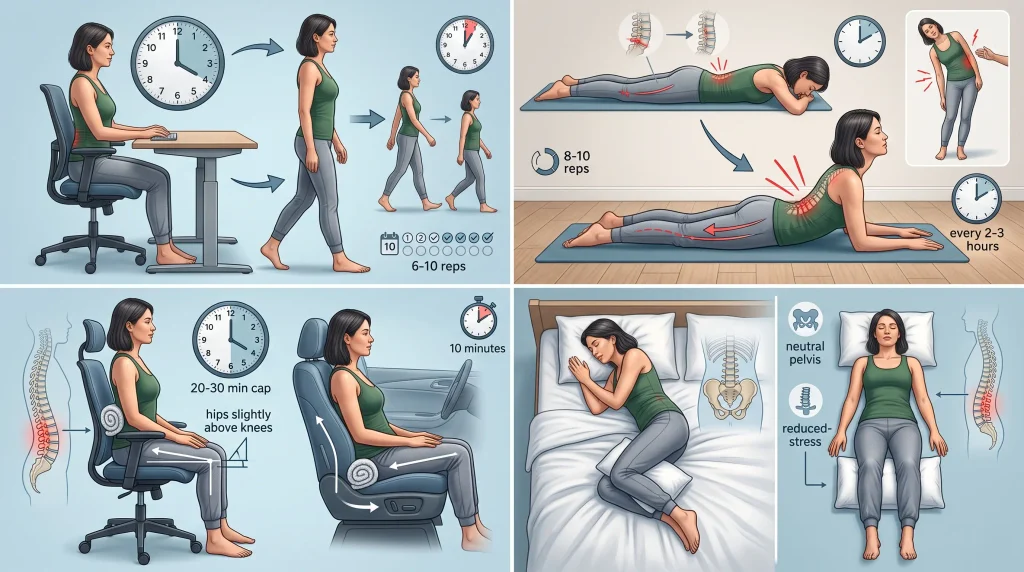
- Movement in small, frequent doses. Get up every 20 to 30 minutes and walk three to five minutes. Six to ten of these beats one long walk: the goal is avoiding sustained positions, not distance.
- Find your directional preference — the movement direction that pulls symptoms out of the leg toward the spine, a response called centralization. For many posterior herniations it is gentle backward bending, lying face down and easing onto the forearms, 8 to 10 repetitions every two to three hours; for others it is a side glide. Judge it by the leg, not the back: more back soreness with a shorter leg symptom is a win. If your trunk has shifted sideways, the antalgic lean of a lateral shift usually needs correcting first.
- Manage sitting deliberately. Cap bouts at 20 to 30 minutes, support the lumbar curve with a rolled towel, and set the seat so hips sit slightly above knees. Driving is the hardest version, since vibration and a reclined pelvis combine — the car seat setup that governs sitting tolerance is worth ten minutes before a long trip.
- Set sleep up properly. Side-lying with a pillow between the knees keeps the pelvis neutral; lying on your back with a pillow under the knees offloads the lumbar spine.
What to do today
- Set a 25-minute timer and walk for three of them every time it goes off, all day.
- Test one direction for five minutes; keep whichever shortens the symptom in your leg.
- Mark today’s symptom endpoint — mid-calf, back of knee, buttock. That is your progress marker.
- Do not spend the day in bed. Short horizontal blocks are fine; a horizontal day is not.
Stage 2 — Movement and unloaded strength (weeks 2–6)
Once leg symptoms retreat above the knee, the work shifts from protection to reconstruction: desensitizing the nerve so it tolerates lengthening again, and rebuilding the hip and trunk contribution that has usually gone missing.
- Nerve gliding moves the nerve through surrounding tissue without stretching it at both ends at once — straightening the knee in sitting as the chin drops toward the chest, then bending the knee as the head lifts. Eight to ten slow repetitions, two or three times daily; if symptoms stay elevated an hour later, reduce range rather than stopping.
- Hip work. Glute bridges 2–3 × 10–12; side-lying hip abduction 2 × 12–15 per side; half-kneeling hip flexor mobility 2 × 30 seconds. A hip that cannot extend or abduct properly forces the lumbar spine to compensate on every step and lift.
- Trunk control without spinal flexion. Dead bug 2–3 × 8 per side; side plank from the knees 3 × 15–20 seconds; bird dog 2–3 × 8 per side. Sit-ups stay out at this stage — not forever — because repeated loaded flexion is the exact mechanism that irritated the annulus.
- Unloaded hinge rehearsal. With a broom handle along the spine touching head, upper back and tailbone, push the hips backward keeping all three contact points, 3 × 8. Everything in Stage 3 is built on this pattern.
- Walking progression to a continuous 20 to 30 minutes, roughly 1.5 to 2.5 km (1 to 1.5 miles). Reciprocal hip movement mobilizes the nerve along its course without the end-range stretch that provokes it.
Stage 3 — Rebuild capacity (weeks 6–12)
This is the stage people skip, and skipping it is why herniations recur. Symptoms are mild by now, which feels like an endpoint and is not.
- Loaded hip hinge, twice weekly. Start elevated — a kettlebell or barbell on blocks so you lift from mid-shin rather than the floor — 3 × 6–8. Lowering block height over successive weeks is the progression, ahead of adding weight. The form errors that cause back pain in the deadlift are the same ones that reload a healing disc.
- Squat pattern. Goblet squats 3 × 8–10, to whatever depth you reach before the lower back tucks under.
- Loaded carries. Suitcase carry with weight in one hand, 4 lengths of 20 to 30 m (22 to 33 yd) per side. Carries train the trunk to resist load with no spinal movement — the exact demand of carrying shopping, a toolbox or a toddler.
- Single-leg strength. Split squats or step-ups 3 × 8 per side; most real lifting happens from a staggered stance.
- Graded occupational rehearsal. If your job means lifting 20 kg (44 lb) boxes from the floor, that exact task needs practicing before your first shift back, not discovering on day one.
The loading rule is simple: in any week increase either load or volume, not both, and keep increases around 5 to 10 percent. Judge the response at 24 hours — soreness gone by next morning is adaptation, while leg symptoms travelling further down mean regress one step and rebuild.
Stage 4 — Full demand and maintenance (week 12 onward)
Full demand means different things for a firefighter, a parent of small children and a recreational lifter, so this stage mirrors what you actually have to do. For impact sport, running returns as a walk-run progression once you can hop comfortably on one leg and carry load without symptom change. For heavy lifting, weight returns at sub-maximal loads from a technically clean position before intensity climbs — the return to heavy lifting after an L5-S1 herniation is a useful template for how conservative that ramp should be. Maintenance decides recurrence: two sessions a week containing a hinge, a carry and anti-rotation trunk work, for about forty minutes in total.
Do’s and don’ts that actually matter for a disc
- Do track where your symptom ends, not how much it hurts. A leg symptom that has moved from calf to buttock is improving even if intensity has barely changed.
- Do brace and hinge at the hip together. Bracing alone, without hip movement, sends load straight to the segment you are protecting.
- Don’t do repeated loaded spinal flexion early. Sit-ups, timed toe touches, rowing with a rounding lumbar spine and heavy kettlebell swings reproduce the pattern that irritates an annular tear.
- Don’t stretch into the leg symptom. A hamstring that feels tight with sciatica is usually a guarded nerve, not a short muscle, and hard stretching pulls on an already sensitized root.
- Don’t collect treatments. Two or three passive sessions to make movement possible is reasonable; six months of appointments with no loading attached is time spent not recovering.
What happens if conservative care stalls?
If leg pain remains genuinely disabling after a fair trial of well-directed conservative care, escalation is reasonable rather than a failure. Two options exist: an epidural steroid injection, which may reduce severe nerve root pain enough to let rehabilitation proceed, and surgery — most often a microdiscectomy — which removes the fragment pressing on the nerve. Both target leg pain from nerve compression, not back pain in general.
The word “fair” is doing real work there. A fair trial means several weeks of a program that actually progressed — staged loading, directional work, hip and trunk rebuilding — not months of appointments and rest. It is common to meet someone at the surgical conversation who never completed anything resembling Stage 2. Guidance on low back pain and sciatica supports considering spinal decompression when non-surgical treatment has not improved pain or function and radiological findings match the symptoms — both conditions, not either alone.
An injection delivers an anti-inflammatory close to the irritated root and can be transformative when pain is too severe to walk, sit or sleep, but it does not change the herniation; its value lies in what you do with the window it opens. Surgery has a different profile: in carefully selected people whose compression matches their symptom distribution, decompression tends to relieve leg pain faster than continued conservative care, with the gap narrowing over the following year. It is far less reliable for back pain without leg symptoms. Progressive neurological deficit or suspected cauda equina compression moves it from elective to urgent.
Putting the plan into practice
Most people with disc-related sciatica improve substantially over six to twelve weeks and never need an operation. Leg pain usually settles before back pain, and numbness lingers longest — a residual patch of altered sensation in the foot does not indicate ongoing compression once strength and reflexes are stable. Treating a herniated disc without surgery is not passive waiting; it is a staged rebuild where each phase has a job and each transition has a test.
Start today: mark where your leg symptom currently ends, set a 25-minute sitting timer, and test one directional movement for five minutes. If that endpoint moves up your leg within two weeks, you are in Stage 2 and the plan is working. If it has not moved at all, get assessed rather than repeat another fortnight of the same thing.
Frequently asked questions
How long does a herniated disc take to heal without surgery?
Most people see meaningful improvement within six to twelve weeks, with the sharpest change in the first month. Full load tolerance takes longer, commonly three to six months, because strength lags well behind pain relief. Numbness can persist after pain has gone and is not a sign healing has stalled.
Can a herniated disc go back in on its own?
Not in the way people picture it, though the outcome is often better than “going back in” implies. Herniated material is broken down and reabsorbed rather than sliding back into the disc, and larger extruded fragments tend to shrink more readily than small contained bulges.
Will lifting weights again damage the disc further?
Loading a healing disc progressively builds tolerance rather than damaging it, provided the movement is a hip hinge rather than a lumbar bend. The risk sits in returning to previous weights before pattern and capacity are rebuilt. Judge each session by where your leg symptoms sit 24 hours later.
Do I need a repeat MRI to confirm the disc has healed?
No, if symptoms and function are improving. Scan appearance correlates poorly with how someone feels, so a repeat MRI in an improving patient almost never changes the plan. Rescanning is justified by new or worsening neurological signs, a changed symptom pattern, or an active surgical decision.
If I have an epidural injection, does that mean surgery is next?
No — an injection is not a step on a conveyor belt toward the operating theatre. It is used to bring severe nerve pain down far enough that rehabilitation becomes possible, and many people who have one never proceed to surgery. What determines the next step is whether the quieter window gets used for rebuilding.
Can I keep working a desk job with a herniated disc?
Usually yes, if you restructure how you sit rather than how many hours you work. Cap sitting bouts at 20 to 30 minutes with a short walk between them, support the lumbar curve, and take calls standing. Staying at work in modified form generally produces better outcomes than a prolonged absence.
Medical disclaimer
This article describes general rehabilitation principles for lumbar disc herniation and cannot replace an individual assessment. Nerve root symptoms need examining in person — strength, reflexes and sensation change the plan, and no article can test them. The staged program here assumes a straightforward disc-related presentation without red flag features; it is not appropriate for suspected fracture, infection, inflammatory disease, cancer or cauda equina compression. Any symptom involving the bladder, bowel or genital area, or weakness that is worsening, needs same-day emergency assessment.
References
- National Institute for Health and Care Excellence. Low back pain and sciatica in over 16s: assessment and management (NG59). 2016, last updated 2020. https://www.nice.org.uk/guidance/ng59
- Hayden JA, Wilson MN, Riley RD, Iles R, Pincus T, Ogilvie R. Individual recovery expectations and prognosis of outcomes in non-specific low back pain: prognostic factor review. Cochrane Database of Systematic Reviews. 2019;(11):CD011284. https://doi.org/10.1002/14651858.CD011284.pub2