
An X-ray shows the bones and alignment of your lumbar spine — but it cannot show discs, nerves, or soft tissues. Common report findings like “disc space narrowing,” “osteophytes,” or “spondylolisthesis” often sound alarming but are frequently age-related changes that do not always cause pain. This guide explains exactly what each phrase on your X-ray report means in plain language, written for patients not clinicians.
An X-ray images bone and alignment only. Discs, nerves, muscles and ligaments are effectively invisible on it, which is why a report can look alarming and still explain very little about your pain. Degenerative findings are also extremely common in people who have no back pain at all — disc degeneration appears in roughly half of pain-free 30-year-olds and in 88% of pain-free 60-year-olds.
Key takeaways
- It is a bone test, not a pain test. A low back pain X-ray shows bone shape, alignment and the gaps between vertebrae. The disc itself, the nerve roots, and the surrounding muscles and ligaments do not appear on it.
- Most findings are also present in people with no pain. Disc degeneration, facet degeneration and mild slips become steadily more common with each decade of life, whether or not a person has symptoms.
- The report describes; it does not diagnose. A radiologist comments on what is visible. Deciding whether any of it explains your pain requires your history and a physical examination.
- Alarming words are usually descriptive. Spondylosis, osteophytes, disc space narrowing and straightening are observations about structure, not predictions about your future.
- Transitional anatomy is worth recording. If sacralization or lumbarization is mentioned, keep a note of it — it changes how spinal levels are numbered for any future injection or surgery.
What does a low back pain X-ray actually show?
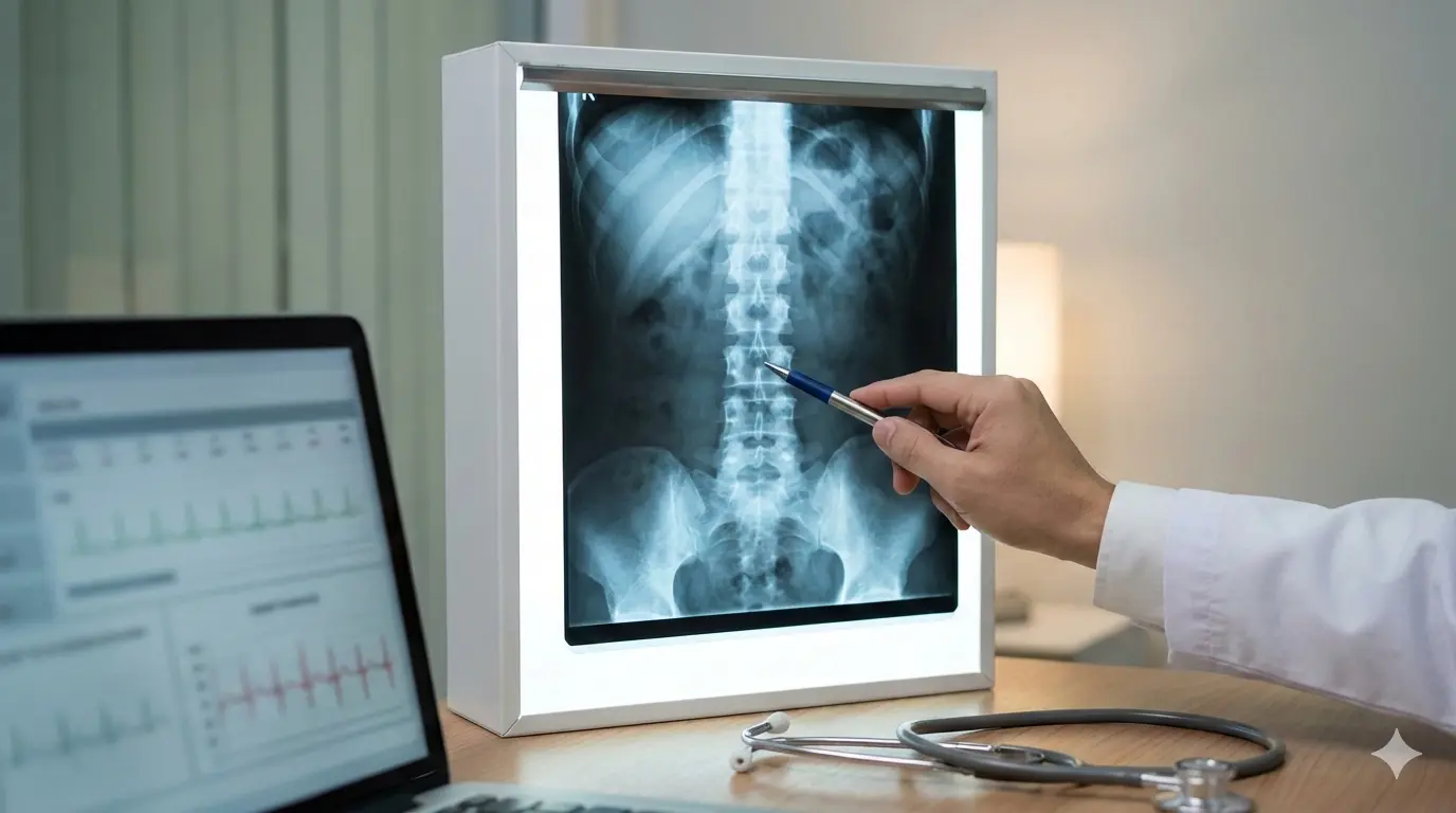
An X-ray is a picture made using a small dose of ionizing radiation. Dense structures—especially bone—block more X-rays and appear brighter/whiter. Less dense structures—muscles, ligaments, discs, and nerves—do not show well and may be essentially invisible. That single fact about how the machine works explains most of the confusion patients have when they read their report.
So an X-ray is best viewed as a “bone and alignment test,” not a “pinpoint the pain source” test.
An X-ray is good at showing:
- Bone shape and bone injury (for example, fractures, compression)
- Alignment and curves (for example, scoliosis)
- Vertebra-to-vertebra position changes (for example, spondylolisthesis)
- Indirect clues about disc height (seen as disc space narrowing)
- Some congenital/anatomic variants (for example, sacralization)
An X-ray is not good at showing:
- Disc bulges, disc herniations, or disc tears (MRI is usually needed)
- Nerve compression (MRI is usually needed)
- Muscles, tendons, and many ligament injuries
- Many early infections, early tumors, or inflammatory changes
Understanding what your report cannot show is just as important as understanding what it can — and your symptoms often tell a clearer story than the images do. That gap—what you want to know vs what the test can show—is why reports can feel unsatisfying.
How many bones are in the low back, and how are they “counted”?
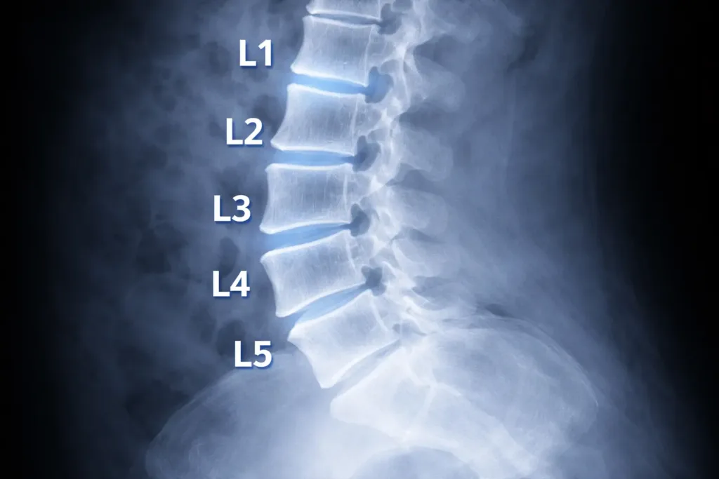
Most people want a straightforward answer: “How many bones do I have here, and what am I looking at?”
The lumbar spine: usually five vertebrae
The lumbar spine typically has five vertebrae: L1, L2, L3, L4, and L5. They sit between the rib-bearing thoracic spine (T12 is just above L1) and the sacrum (S1 is just below L5).
On imaging, L5–S1 is the most discussed level because it is the junction between the mobile lumbar spine and the fixed pelvis.
The sacrum: usually five fused segments
Below L5 is the sacrum, typically made of five fused vertebrae (S1 to S5). It forms the back wall of the pelvis and connects to the hip bones at the sacroiliac joints.
The coccyx: usually 3–5 small fused bones
Below the sacrum is the coccyx (tailbone), commonly 3–5 small segments fused to varying degrees.
Why counting can be tricky
Counting levels is not always as simple as “five lumbar bones, then sacrum.” Some people have an anatomical variation called a lumbosacral transitional vertebra, where the border between lumbar spine and sacrum is blurred. This includes:
- Sacralization (L5 behaves more like part of the sacrum)
- Lumbarization (S1 behaves more like an extra lumbar vertebra)
These variants can change how a radiologist numbers the levels and can matter later if you ever need an injection, procedure, or surgery—because the “level” must be identified correctly.
What happens during a low back pain X-ray?
The appointment is usually short. Expect to change into a gown, remove anything metallic near your waist, and hold two or three positions while the images are taken. The imaging itself often takes only a few minutes; positioning and paperwork take longer. You will not feel anything during the exposure, and no injection or contrast dye is involved.
What you may be asked to do
- Change into a gown: Metal on clothing can obscure the image (belts, zippers, hooks).
- Remove nearby items: Coins, keys, jewelry, or anything metallic near the abdomen/pelvis.
- Stand or lie down: Depending on the requested views and your comfort.
- Hold your breath briefly: A short breath-hold reduces motion blur.
- Turn slightly for angles: If the team is capturing oblique or spot views.
Common views in a lumbar X-ray series
Radiology practices vary, but a typical X-ray series may include:
- AP view: A front-to-back image that helps assess overall alignment and side-to-side symmetry.
- Lateral view: A side image that shows the curve (lordosis), disc spaces, and slips more clearly.
- L5–S1 spot view: Sometimes used to better visualize the lowest disc space through the pelvis.
- Oblique views: Less common in routine care now; historically used for pars defects (the “Scotty dog” concept).
- Flexion–extension views: Sometimes requested if instability is suspected; you bend forward and backward to see whether segments translate abnormally.
If your report references “standing” images, that detail can matter because gravity may reveal alignment issues that are less obvious when lying down.
What your report is really doing: describing, not diagnosing
Radiology reports are descriptive summaries of visible anatomy. They are often written to answer a narrow clinical question (for example, “Is there a fracture?” or “Is there a slip?”).
A helpful patient perspective shift:
- The report describes what is visible on an X-ray.
- Your clinician integrates the report with your symptoms, timeline, and physical exam.
That is why two people with similar “degenerative changes” on an X-ray can feel very different: the image is not measuring pain.
What do the phrases on a low back pain X-ray report actually mean?
Most report phrases fall into three groups: statements that nothing urgent was found, descriptions of age-related change at the bones and joints, and notes about alignment or anatomical variation. Very few of them name a cause of pain. The decoder below translates the terms that appear most often, in the order you are most likely to meet them.
“No acute osseous abnormality”
This generally means “no new bone problem was seen,” such as no fresh fracture or destructive bone lesion.
It does not mean “no reason for pain exists.”
“Degenerative changes,” “spondylosis,” or “osteoarthritic changes”
These phrases describe wear-and-adaptation patterns at the bony margins and joints—often small bone spurs (osteophytes) and mild joint changes.
These findings are common and tend to increase with age. They can be relevant, but they are not automatically the cause of pain.
“Osteophytes”
Osteophytes are bone spurs. On an X-ray they may be described as “anterior osteophytes” or “marginal osteophytes.” Many are incidental. Their significance depends on size, location, and whether they plausibly match your symptom pattern.
“Disc space narrowing”
An X-ray cannot see the disc directly; it sees the space between vertebrae. Narrowing suggests reduced disc height, often from disc dehydration and long-term change.
Disc space narrowing does not confirm a disc herniation and does not measure nerve compression.
“Facet arthropathy”
Facet joints are small joints at the back of the spine that guide movement. “Facet arthropathy” means arthritis-like change in those joints.
On an X-ray, facet changes can be under-reported because facets are harder to see well than on CT. If this term appears, it generally means the changes were visible enough to comment on.
“Spondylolisthesis,” “anterolisthesis,” or “retrolisthesis”
These terms describe a vertebra slipping forward (anterolisthesis) or backward (retrolisthesis) relative to the one below.
If the report gives a “grade,” it is referring to the degree of slip (Grade 1 is mild). Sometimes the report also mentions whether it appears stable or suggests flexion–extension views.
“Loss of lordosis” or “straightening”
The lumbar spine normally has a gentle inward curve (lordosis). “Straightening” can reflect muscle guarding, positioning during the X-ray, or day-to-day posture.
It is frequently reversible and should not be treated as a permanent structural defect based on wording alone.
“Scoliosis”
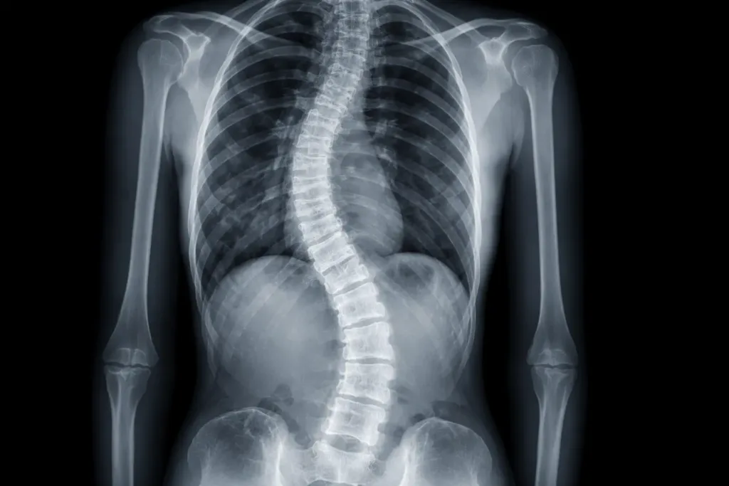
Scoliosis is a sideways curve. A report may describe the direction (left or right), location, and sometimes angle. Some curves are structural; others are “functional” and appear during pain episodes or protective muscle tightening.
“Transitional anatomy,” “sacralization,” or “lumbarization”
These terms matter. They describe a variant where the lowest lumbar vertebra (L5) or the top sacral segment (S1) has features of the other region.
If your report mentions transitional anatomy, it is worth ensuring your clinician notes it for future reference.
Sacralization: the missed finding patients should understand
Sacralization is commonly under-explained in routine conversations, yet it can change how your X-ray is interpreted and how levels are labeled.
What sacralization means
Sacralization is when L5 is partially or completely fused to the sacrum, or when L5 has an enlarged transverse process that forms a joint or fusion with the sacrum or pelvic bone.
In plain terms: the “lowest lumbar bone” behaves more like part of the sacrum.
How it shows up on an X-ray report
Radiologists may write:
- “Lumbosacral transitional vertebra”
- “Sacralization of L5”
- “Pseudoarticulation of the transverse process”
- “Bertolotti syndrome” (used when the transitional segment is suspected to be pain-related)
Why it matters to you
Sacralization matters for three practical reasons:
- Level numbering can be confusing
With sacralization, what one clinician calls “L5” another might label differently depending on how they count. This matters if someone ever plans an injection, procedure, or surgery. Correct level identification may require additional counting methods or comparison imaging. - Load and motion can shift
When one segment is more fused or behaves differently, motion and load can shift to the level above. That can influence where changes appear on an X-ray (often above the transitional segment). - It can be linked to symptoms in some people, but not in all
Many people with sacralization have no symptoms. In others, the transitional joint can become irritated and contribute to back or buttock pain. The key point: sacralization is context, not a standalone diagnosis.
Lumbarization: the mirror image
Lumbarization is when the top sacral segment (S1) looks and behaves more like an extra lumbar vertebra. The same cautions apply: numbering can differ, and any relevance must be tied to symptoms.
How radiologists decide what to mention (and what they often do not)
Understanding what drives the report helps you read it more calmly.
Reports prioritize what is reliably visible
Because discs and nerves are not well seen, X-ray reports tend to emphasize:
- Alignment and curvature
- Bone integrity (fracture vs no fracture)
- Disc space height (indirect)
- Visible joint changes
- Congenital variants (sometimes)
Many “normal variations” are not listed
Reports may not mention small asymmetries or minor variants if they are not considered clinically significant. That can be frustrating, but it is a common reporting practice.
Template language is common
Some reports use structured templates, which can make wording feel generic. The useful part is the specific findings and the impression, not the phrasing style.
Why doesn’t my X-ray match how I feel?
Because the findings on your film are common in people who feel perfectly well. A systematic review of spinal imaging in adults with no back pain at all found disc degeneration in 37% of 20-year-olds rising to 96% of 80-year-olds, and facet degeneration in 4% of 20-year-olds rising to 83% of 80-year-olds. Structure and symptoms often do not track together.
From a patient perspective, the biggest confusion is mismatch:
- “My X-ray looks bad, but I feel okay.”
- “My X-ray looks normal, but I feel terrible.”
Both are common. Reasons include:
- Pain can come from discs, nerves, muscles, and ligaments—structures that an X-ray does not show well.
- Some X-ray findings (like mild degenerative changes) are common even in people without pain.
- Pain can be influenced by inflammation and nerve sensitivity, which are physiologic processes not visualized on X-ray.
A practical mindset is: an X-ray is excellent at ruling in or ruling out certain categories of problems, but it is limited for explaining pain intensity.
Do I need an MRI instead of an X-ray?
Usually not, and rarely as the first test. An X-ray answers bone questions — fracture, alignment, slip, transitional anatomy — quickly and cheaply. MRI becomes the better choice when symptoms travel down the leg, when weakness or numbness is present, when pain persists despite reasonable conservative care, or when a procedure is being planned. The table below sets out what each test can and cannot answer.
| Test | What it is best at | What it often cannot answer | Typical patient scenario |
|---|---|---|---|
| X-ray | Bones, alignment, fractures, transitional anatomy, slips | Disc herniation, nerve compression, most soft tissues | Trauma concerns, suspected instability, baseline alignment questions |
| MRI | Discs, nerves, soft tissue, inflammation | Some bony detail; many incidental findings | Persistent leg symptoms, suspected nerve compression, pre-procedure planning |
| CT | High-detail bone imaging | Higher radiation; less soft tissue detail than MRI | Complex fractures, detailed bony assessment when MRI is not suitable |
Is the radiation from a lumbar X-ray something to worry about?
For a healthy adult, a single lumbar series carries a small radiation dose, and the associated risk is generally considered low. That is not a reason to be casual about it. The practical concern is rarely one X-ray — it is repeated imaging ordered over months that is unlikely to change any treatment decision, which is where avoidable exposure accumulates.
Two practical points follow from that. Repeat imaging is worth questioning if nothing about your symptoms has changed, since a second film of an unchanged back rarely alters treatment. And if pregnancy is possible, say so before the images are taken so the team can adjust shielding or reconsider the test.
How do I get the most out of my X-ray appointment?
These patient-centered steps can make the result more useful later:
- Ask whether images are standing or lying down; standing images can better reflect alignment under load.
- If you have prior imaging, ensure it is available for comparison; “no change from prior” can be meaningful.
- Request access to the actual images (portal download or disc) in addition to the report.
- If your report mentions transitional anatomy such as sacralization, keep a note of that for future procedures so level labeling stays consistent.
👨⚕️ Dr. Arora’s Clinical Note:
A common mistake I see is treating the X-ray appointment like a formality and then expecting the report to “explain the pain.” Here’s the practical upgrade that makes imaging more usable: mark the symptom, not just the spine. Before the lateral image, ask the technologist if they can place a small skin marker (they often use a tiny BB-style marker) over your single most painful point (right vs left, near the beltline vs closer to the tailbone). It doesn’t diagnose anything—but it helps your clinician correlate whether the pain behaves more like facet/SI-region irritation vs midline disc-related patterns, especially when the written report is generic.
Counter-intuitive physio tip: if your report says “loss of lordosis/straightening,” don’t rush to aggressive stretching to “restore the curve.” That phrase is often guarding + positioning, and stretching into pain can increase protective spasm. A better first step is 2–3 days of gentle walking + relaxed diaphragmatic breathing + pain-free hip-hinge practice (micro-movements) to downshift the guarding—then reassess.
Red flags: when imaging should not delay urgent evaluation
A small number of symptoms mean the priority is assessment, not imaging. These features raise the possibility of nerve compression, infection, fracture or cancer affecting the spine — problems where delay changes the outcome. If any of the following apply to you, arrange urgent medical review rather than waiting for a scan appointment or for a report to arrive.
- New or progressive leg weakness. A foot that catches on steps or slaps the floor (foot drop), or a leg that gives way on stairs. Weakness that is worsening suggests a nerve root is losing function rather than simply being irritated, and the window for protecting that nerve is limited.
- Loss of bowel or bladder control, or numbness in the saddle area. Difficulty starting or stopping urination, new incontinence, or numbness where you would contact a saddle. This combination can indicate cauda equina compression, which is treated as a surgical emergency and should be assessed the same day.
- Fever, night sweats or unexplained chills with severe back pain. Infection of a disc, a vertebra or the space around the spinal cord can present as back pain before anything else appears, and plain films are frequently normal in the early stages of spinal infection.
- A history of cancer with new, persistent back pain. The spine is a common site for secondary spread. Pain that is unrelenting, wakes you at night, or is unrelated to position deserves prompt review, because an X-ray can miss early bone involvement.
- Severe pain after significant trauma — or after minor trauma if fracture risk is high. A fall from standing height can fracture a vertebra in someone with osteoporosis or on long-term corticosteroids. This is one situation where an X-ray is genuinely the right first test and should not be postponed.
- Unexplained weight loss alongside back pain that does not settle. A nonspecific signal on its own, but combined with persistent pain it justifies looking beyond the lumbar spine rather than repeating imaging of it.
An X-ray can be part of evaluation, but these features require clinician assessment regardless of imaging.
What to ask after you receive your report
If you only received a report with no explanation, focus on questions that connect the X-ray to decisions:
- “Is there any sign of fracture, destructive bone change, or a significant slip?”
- “Do any alignment findings on the X-ray match my symptom pattern?”
- “Do I have a transitional vertebra such as sacralization, and does it affect level numbering?”
- “Should these X-ray findings be considered stable, or is follow-up imaging ever needed?”
- “If symptoms are mainly leg-related, does MRI add information that the X-ray cannot provide?”
A report cheat sheet for common findings
| Finding on X-ray | What it often reflects | Why patients misread it |
|---|---|---|
| Mild spondylosis / degenerative change | Normal wear/adaptation over time | Assumed to equal damage or inevitability |
| Disc space narrowing | Reduced disc height | Assumed to prove herniation or nerve pinch |
| Osteophytes | Bone spurs at margins | Assumed to be “the cause” by themselves |
| Grade 1 anterolisthesis | Mild forward slip | Assumed to require surgery immediately |
| Loss of lordosis | Guarding/positioning | Assumed to be permanent deformity |
| Transitional vertebra / sacralization | Variant anatomy at L5–S1 | Often not explained; can confuse level labeling |
FAQs about a low back pain X-ray
Does a low back pain X-ray show a disc bulge or herniation?
An X-ray does not directly show disc bulges or herniations. It may show disc space height changes, but MRI is typically used to visualize discs and nerves.
What does “no acute abnormality” mean on an X-ray?
It usually means no new fracture or destructive bone problem was seen. It does not rule out disc, nerve, or soft-tissue causes of symptoms.
If my X-ray shows degeneration, does that mean my back is “wearing out”?
Degenerative changes on an X-ray often reflect common, gradual adaptation. They do not automatically predict severe pain or disability.
Can an X-ray diagnose spinal stenosis?
X-ray can show indirect clues (like arthritis or a slip), but it cannot measure spinal canal narrowing well. MRI is typically used to evaluate stenosis.
What is sacralization, and why is it mentioned on my report?
Sacralization is a lumbosacral transitional vertebra where L5 partly or fully joins the sacrum. It matters because it can affect level numbering and sometimes shifts load to the level above.
What is lumbarization?
Lumbarization is when the top sacral segment behaves more like an extra lumbar vertebra. Like sacralization, it can affect numbering and interpretation.
Why did my clinician order an X-ray instead of MRI?
An X-ray is fast, widely available, and useful for checking bone integrity and alignment. MRI is usually reserved for nerve symptoms, persistent problems, or pre-procedure planning.
Should I worry about radiation from an X-ray?
For most adults, the risk from a single lumbar series is low. The main safety principle is to avoid unnecessary repeat imaging.
Can an X-ray miss a fracture?
Yes. Some fractures can be subtle, and visibility depends on views and bone quality. If suspicion is high, clinicians may consider CT or MRI.
What should I keep from my imaging visit: the report or the images?
Ideally both. The report is a summary; the images allow comparison over time and can be valuable for second opinions—especially if your X-ray notes sacralization or other anatomy that affects level labeling.
For a complete overview of all lower back pain causes and treatments, see our Lower Back Pain Complete Guide.
The bottom line
A low back pain X-ray is a useful test asked to do one job well: check the bones and the way they line up. Read it that way and most of the alarm drains out of the wording. If the findings are degenerative, treat them as context rather than a verdict — they are present in most pain-free adults over 50 too. Judge your progress by what your back lets you do, such as walking distance, sitting tolerance and getting out of a car, rather than by the adjectives on a page. Then bring the report to whoever is guiding your rehabilitation and ask the only question that changes anything: which of these findings, if any, alters the plan? For readers who want the clinician-level source, the Academy of Orthopaedic Physical Therapy’s clinical practice guideline on interventions for acute and chronic low back pain is the document physical therapists work from.
References
- Brinjikji W, Luetmer PH, Comstock B, et al. Systematic literature review of imaging features of spinal degeneration in asymptomatic populations. AJNR Am J Neuroradiol. 2015;36(4):811–816. doi:10.3174/ajnr.A4173. Full text
- George SZ, Fritz JM, Silfies SP, et al; Academy of Orthopaedic Physical Therapy, American Physical Therapy Association. Interventions for the Management of Acute and Chronic Low Back Pain: Revision 2021. J Orthop Sports Phys Ther. 2021;51(11). doi:10.2519/jospt.2021.0304. Guideline summary