
Your MRI report came back and somewhere in the findings is a line about Modic changes. It sounds ominous, nobody explained it, and the internet is not helping. Here is the short version before the detail: this is a common degenerative finding, it is not cancer, it is not a fracture, and on its own it does not decide how much pain you will have or what treatment you need.
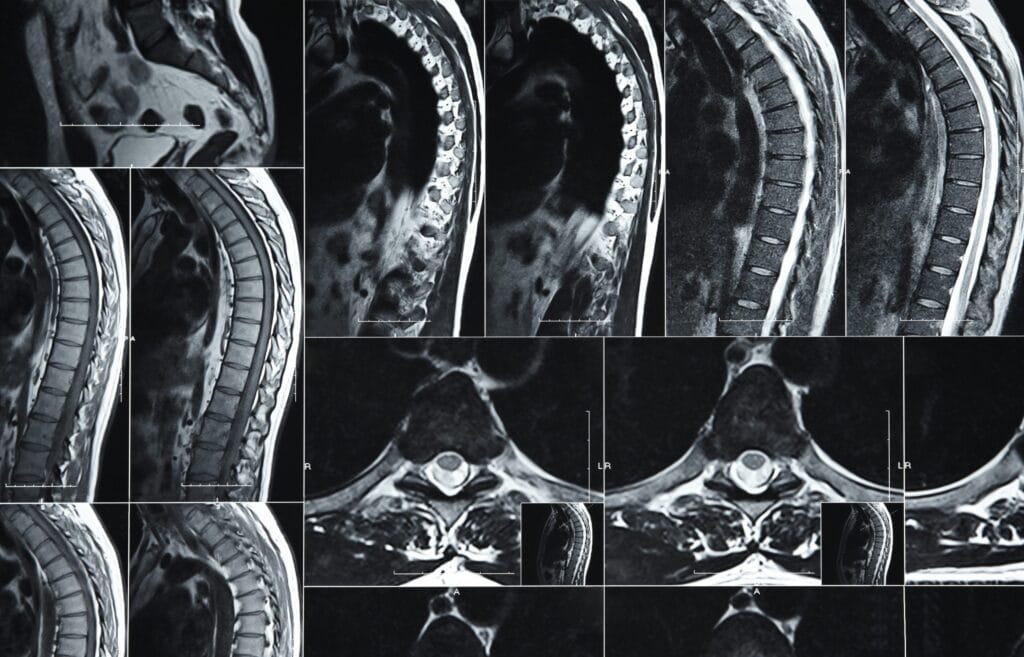
Modic changes on MRI are signal changes in the bone marrow of a vertebra next to the disc, graded Type 1, 2 or 3 depending on what the bone is doing. Type 1 reflects active inflammation and swelling, Type 2 reflects fatty replacement, and Type 3 reflects hardened bone. They indicate wear and irritation at the endplate — not a dangerous disease — and many people who have them are pain-free.
Key takeaways
- The finding describes bone, not disc. Modic changes sit in the vertebral bone marrow beside the endplate, which is why they behave differently from a disc bulge.
- Type matters more than the word itself. Type 1 carries the strongest association with pain; Type 2 is more settled; Type 3 is rarely the pain driver.
- A finding is not a diagnosis. The change only counts clinically if the level on the scan matches the level that reproduces your symptoms on examination.
- Treatment starts conservatively and usually stays there. Load management, hip-driven movement retraining and graded activity resolve or substantially improve most cases.
- One situation needs urgency. Type 1 appearances can occasionally overlap with spinal infection, so fever, night sweats or recent spinal procedures change the picture entirely.
What exactly are Modic changes?
They are alterations in the bone marrow of a vertebra, immediately adjacent to the vertebral endplate — the flat surface where the bone meets the intervertebral disc. Radiologists grade them into three types based on how the marrow behaves on different MRI sequences. The finding tells you that this specific bone-and-disc interface has been under mechanical stress and has responded biologically.
The name comes from Dr. Michael Modic, the radiologist who first classified the pattern. That is all the term means — a descriptive label, not a disease name. It is closer in spirit to a report noting gray hair than to a report noting a tumor: a marker of change over time in a structure that takes load every day of your life.
What do Type 1, Type 2 and Type 3 actually mean?
The three types describe three different biological states in the same location. Type 1 is active inflammation with fluid in the marrow. Type 2 is fatty replacement, generally a more settled and chronic state. Type 3 is sclerosis, where the bone has become dense and hardened. They are stages rather than severities, and one type can convert into another over months to years.
| Type | How it looks on MRI | What the bone is doing | Relationship to pain | Typical course |
|---|---|---|---|---|
| Type 1 | Dark on T1, bright on T2 | Marrow edema — fluid, inflammation, active irritation | Most consistently linked to symptoms of the three types | Often converts to Type 2 over months to years as inflammation settles |
| Type 2 | Bright on T1, bright on T2 | Fatty replacement of marrow — a more chronic, settled state | Can be associated with pain, but less strongly than Type 1 | Usually stable; may persist indefinitely without symptoms |
| Type 3 | Dark on T1, dark on T2 | Sclerosis — dense, hardened bone | Least associated with active pain | Represents long-standing change; rarely the treatment target |
Should I be worried if my report says Modic changes?
In almost all cases, no. This is a degenerative finding, not a sinister one. It does not indicate cancer, does not mean your spine is unstable, and does not put you on a track toward surgery. Many people carry these changes with no back pain at all, which is exactly why the scan cannot be read in isolation from what your body is actually doing.
That said, the association with pain is real and worth understanding honestly. A systematic review and meta-analysis by Herlin and colleagues in PLOS ONE found that in studies using provocative discography, Type 1 changes carried a markedly higher odds of concordant pain than no Modic changes, with Type 2 showing a weaker association. The authors also noted substantial variation between studies — which is the honest summary: a meaningful signal, not a certainty.
The practical translation is straightforward. If your pain is deep, central, worse with sustained sitting and forward bending, and your MRI shows Type 1 changes at a level that hurts when examined, the finding is probably relevant. If your pain is one-sided, worse with extension, and the changes sit two levels away, it probably is not.
Why do Modic changes develop in the first place?
They develop where the endplate has taken more mechanical stress than it could comfortably absorb, and the bone has responded with inflammation, then remodeling. The most common driver is disc degeneration: as a disc loses height and water content, it stops distributing load evenly, and the endplate above and below starts absorbing force it was never designed to take alone.
- Disc height loss and dehydration. A flattened disc transmits load in concentrated points rather than spreading it, which is the single most common setup for endplate change.
- Repetitive microtrauma. Years of loaded forward bending — lifting toddlers, moving boxes, rowing with a rounded back — creates cumulative endplate stress rather than one identifiable injury.
- Prolonged static loading. Long seated hours compress the anterior column continuously, with none of the load variation that walking provides.
- Previous endplate injury. A compression injury that healed with an uneven surface concentrates stress at that point for years afterward.
- Low-grade infection. Uncommon, and debated, but bacterial involvement of the disc has been proposed as one contributor to Type 1 appearances in some cases.
When does a Modic finding need urgent assessment?
Modic changes themselves are not an emergency. The reason to pay attention is that Type 1 appearances — marrow fluid and inflammation — can look similar to early spinal infection on imaging, and infection is a genuine emergency. The following features should prompt medical assessment rather than watchful waiting, because each one changes the differential.
- Fever, chills, night sweats or feeling systemically unwell alongside back pain. This combination raises the possibility of discitis or vertebral osteomyelitis, which requires urgent blood tests and specialist review rather than physical therapy.
- Recent spinal injection, spinal surgery, or a bloodstream infection in the past few months. These are the routes by which infection reaches the spine, and they meaningfully raise the index of suspicion for any new Type 1 pattern.
- Back pain that is worse at night, wakes you, and does not ease with any position change. Mechanical endplate pain nearly always responds to unloading; pain that ignores position deserves a wider workup.
- New numbness in the saddle region, or loss of bladder or bowel control. This suggests compression of the nerve bundle at the base of the spinal cord and requires same-day emergency assessment.
- Progressive leg weakness, unexplained weight loss, or a history of cancer. Each points away from a straightforward degenerative explanation and toward a diagnosis that must be excluded first.
What treatment helps when Modic changes are causing pain?
Treatment targets the mechanical situation that produced the change, not the MRI appearance. That means restoring tolerance to compressive load, shifting bending demand into the hips, and rebuilding capacity gradually. Medication and injections can create a window in which that work becomes possible. A targeted nerve procedure is reserved for a small, well-defined group who have already completed genuine conservative care.
Because the pain mechanism here is the same one described in vertebrogenic low back pain, the rehabilitation logic is identical: interrupt sustained compression, load the spine progressively rather than protect it indefinitely, and judge progress by how the back feels the next morning rather than by how it feels mid-session.
For the minority whose pain persists beyond six months of well-executed conservative care, basivertebral nerve ablation targets the nerve carrying the pain signal from inside the vertebral body. Candidate selection specifically requires documented Type 1 or Type 2 changes on MRI along with a matching clinical picture, as set out in the StatPearls review of basivertebral nerve ablation. It is a narrow intervention for a narrow group.
Quick check: does this finding explain your pain?
1. Is your pain deep and central rather than one-sided?
2. Does sustained sitting or standing still make it worse, and lying flat make it better?
3. Does forward bending aggravate it more than leaning back?
4. Is the pain mostly in the back rather than traveling down the leg?
5. Does the level named in your report match where you actually feel it?Mostly yes: the finding is likely relevant, and endplate-focused rehabilitation is a sensible starting point. Mostly no: the changes may be incidental, and the real driver is worth reassessing before you build a treatment plan around the scan.
Can Modic changes go away?
The MRI appearance rarely disappears completely, but it does change. Type 1 changes commonly convert to Type 2 as active inflammation settles into fatty marrow, which is generally accompanied by an improvement in symptoms. The more useful question is not whether the scan will normalize but whether your tolerance for sitting, walking and lifting improves — and that reliably can improve, even when the imaging looks the same.
This is worth holding onto if you are inclined to re-scan every few months. Repeat imaging in the absence of new red-flag features rarely changes management and frequently increases anxiety. Function is the outcome that matters.
The bottom line
Modic changes on MRI are a description of how one part of your spine has aged and responded to load. In the right clinical context they identify a specific, treatable pain source and point toward a targeted plan. In the wrong context they are an incidental finding that has been given far more weight than it deserves. Knowing which situation you are in is the whole task — and it takes an examination, not a report.
Frequently asked questions
Are Modic changes always painful? No. Type 2 and Type 3 changes are frequently found in people with no symptoms, and even Type 1 changes are not universally painful. The finding raises the probability that the endplate is involved; it does not confirm it.
Is Type 1 worse than Type 2? Not worse in terms of damage, but more likely to be actively symptomatic. Type 1 represents inflammation in progress, which is why it correlates more strongly with pain and why it also has more potential to improve.
Do Modic changes mean I will need surgery? Almost never. The overwhelming majority of cases are managed with conservative care, and where that fails, minimally invasive options are considered well before any discussion of fusion.
Should I avoid the gym? No, but choose carefully. Repeated loaded spinal flexion — sit-ups, heavy rounded-back deadlifts, long rowing sessions with a collapsed posture — tends to aggravate endplate pain, while walking, hip hinge patterns and neutral-spine strength work are generally well tolerated.
Is there a specific exercise for Modic changes? There is no single exercise that treats the imaging finding. What changes symptoms is reducing total daily compressive load, improving hip mobility and strength so the lumbar spine bends less, and increasing loading tolerance in steady increments.
Why did my previous MRI not mention them? Modic changes develop over years, and reporting also varies between radiologists and scanner sequences. A new mention does not necessarily mean the change appeared since your last scan.
How common are they? Common in adults with chronic low back pain and degenerative disc disease, and present in a meaningful proportion of people without symptoms. Reported prevalence varies widely between study populations, which is part of why the finding must be interpreted clinically.
References
- Herlin C, Kjaer P, Espeland A, et al. Modic changes — their associations with low back pain and activity limitation: a systematic literature review and meta-analysis. PLOS ONE. 2018;13(8):e0200677. doi:10.1371/journal.pone.0200677 (PMID: 30073201)
- Tieppo Francio V, Sayed D. Basivertebral Nerve Ablation. In: StatPearls [Internet]. Treasure Island (FL): StatPearls Publishing; 2023. NCBI Bookshelf NBK572127
With 20+ years of clinical experience in spine, joint, and pain management, Dr. Vivek ensures every article reflects accurate, trustworthy, and up-to-date health advice.
Learn more about Dr. Vivek’s qualifications & journey →