Expert Review: This article includes clinical insights from Dr. Vivek Arora, a physiotherapist with 20+ years of experience.
Pain that sits low and off to one side of the pelvis — below the belt line, near the small dimple at the back — is one of the most misattributed complaints in musculoskeletal care. Some of it is lumbar, some is hip, and a genuine share is sacroiliac joint dysfunction: a mechanical pain problem from the joint where the base of the spine meets the pelvis. The label has collected a lot of folklore, most of it involving a joint that supposedly slips out and needs putting back. It barely moves, and the problem is almost never a bone in the wrong place.
Key takeaways
- The joint does not “go out of place.” It moves a few millimeters. Pain comes from ligaments, capsule and joint surfaces irritated under load.
- Location is informative. One-sided pain within a thumb’s width of the back dimple is the most suggestive single feature.
- Single-leg loading is the giveaway. Stairs, stepping out of a car, rolling in bed and dressing while standing provoke it more than sitting.
- No single test is reliable. Clinicians use a cluster of provocation tests and look for three or more positives — a probability, not a verdict.
- One pattern is not mechanical. Morning stiffness over 30 minutes, waking in the second half of the night, and pain that eases with movement may indicate inflammatory sacroiliitis, which needs medical assessment.
What is sacroiliac joint dysfunction, and does the joint really go out of place?
Sacroiliac joint dysfunction is pain from the joint between the sacrum — the triangular bone at the base of the spine — and the ilium, the flared bone of the pelvis. It is a load-related mechanical problem, not a displacement: the joint has only a few millimeters of motion, so it cannot slip out of position and be pushed back in.
Its job is transferring force. Every step sends the weight of your head, arms and trunk down the spine into the sacrum, across the pelvis, and out into whichever leg is on the ground. No muscle moves this joint; it is wedged in place by interlocking surfaces and thick ligaments, with muscles crossing the region adding compression on top — gluteus maximus, the opposite latissimus dorsi, the deep hip rotators, the pelvic floor, the abdominal wall. When that muscular contribution falls off through weakness, post-childbirth inhibition or fatigue, ligaments and capsule absorb shear they were meant to share. Nothing is misaligned — which makes “dysfunction” an unfortunate word for a joint asked to tolerate more asymmetric load than it can.
Three related beliefs are worth dropping. The joint does not go out and back in — week to week, what changes is guarding and pain sensitivity. A pelvis that palpates as “higher” is a poor guide, since small asymmetries are unreliably detected and common in people with no symptoms. And prolonged rest backfires: less activity means less muscular compression, so tolerance falls further.
What does sacroiliac joint pain actually feel like?
SI joint pain is usually one-sided, sits below the belt line near the back dimple, and feels deep and localized rather than shooting. People often point to it with one finger. It can spread into the buttock and back of the thigh, but rarely past the knee, and loading one leg at a time provokes it more than bending.
The landmark worth knowing is the posterior superior iliac spine, the bony bump creating the indentation either side of the lower back. Pain centered within about 2.5 cm (1 inch) of it is a useful pointer, which is why pain around the lower back dimples is a location clue rather than vague back pain. Relief comes from symmetry — lying flat, weight evenly split, level walking.
Quick check: does this look like an SI joint pattern?
- Can you cover the worst of the pain with one hand, below the belt line, on one side?
- Is stepping up onto a stair or curb clearly worse on that side?
- Does rolling over in bed make you brace or wake you?
- Does bending forward bother you less than standing on one leg?
Mostly yes: an SI pattern is plausible and a movement assessment is the next step. Mostly no — especially if bending and sitting are worst — the lumbar spine is the likelier source.
Who tends to get sacroiliac joint dysfunction?
Five histories load this joint far more than average: pregnancy and the postpartum year, a fall onto one buttock, generalized hypermobility, a marked leg-length difference, and previous lumbar fusion. What links them is a step change in the one-sided shear the joint absorbs, or a drop in how well muscle shares it.
In pregnancy, ligament laxity rises and the center of mass shifts forward, so stability falls exactly as walking demand climbs. Symptoms often outlast delivery — a common thread in back and pelvic pain still present a year after a caesarean, where deep abdominal and gluteal muscles were never retrained. A fall onto one buttock strains the posterior ligaments directly. In hypermobility, elastic ligaments lock the joint less and muscle must do more — which is why this group, often flexible and sometimes very fit, is surprised to need stability work rather than stretching. A marked leg-length difference alters the size and timing of force reaching each joint thousands of times a day, and after lumbar fusion, load once shared across the fused segments has to go somewhere.
How do clinicians test for it?
No single physical test confirms sacroiliac joint pain. Clinicians use a cluster of pain-provocation tests, each loading the joint differently, and read the result as a group — three or more positives out of five is the threshold usually treated as meaningful. The tests reproduce familiar pain rather than measure movement, because measuring millimeters of motion through skin is not realistic.
- Distraction — outward, backward pressure on both front pelvic bones, gapping the front of the joint.
- Thigh thrust — hip bent to about 90 degrees, force directed down the femur to shear the joint surface.
- Compression — side-lying, downward pressure through the upper pelvic rim.
- Sacral thrust — face down, pressure through the center of the sacrum.
- Gaenslen’s test — one hip pulled into deep flexion while the other extends off the table edge, twisting the halves of the pelvis oppositely.
Two caveats get skipped far too often. The cluster is a probability tool that raises or lowers suspicion; its accuracy should not be oversold. And it loses usefulness when signs point to a lumbar disc — particularly when repeated movements make leg symptoms retreat toward the spine. Palpation alone is weak: joint-line tenderness is common in people with no SI problem.
Could it be something other than the SI joint?
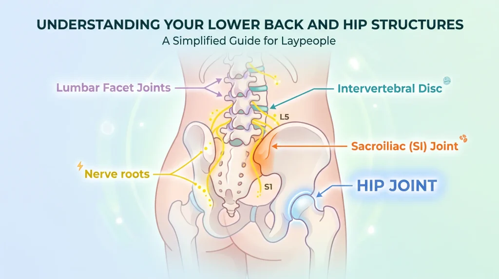
Yes — four alternatives cause most of the confusion. Lumbar facet joint pain, a lumbar disc with nerve root irritation, hip joint pathology and inflammatory sacroiliitis all produce pain around the buttock and lower back. They separate reasonably well on where the pain sits, what provokes it, what relieves it, and when it peaks.
| Source | Where it sits | Worse with | Better with | Distinguishing clue |
|---|---|---|---|---|
| SI joint | One side, below the belt line, near the back dimple | Stairs, single-leg standing, rolling in bed | Lying flat, even weight-bearing | Coverable with one hand; rarely past the knee |
| Lumbar facet | Higher, at or above the belt line, nearer the midline | Leaning back, twisting, standing still | Sitting, bending forward | Extension and rotation aggravate, not single-leg load |
| Disc / nerve root | Back plus a defined band down the leg, often below the knee | Sitting, bending, coughing | Standing, walking | Numbness, tingling or weakness in a set distribution |
| Hip joint | Groin, front of thigh, deep lateral buttock | Deep squatting, pivoting, putting on socks | Avoiding end-range rotation | Reduced hip rotation; groin pain is the strongest pointer |
| Inflammatory sacroiliitis | Alternating or both-sided buttock pain, often deep | Rest, inactivity, second half of the night | Movement, exercise, a hot shower | Morning stiffness over 30 minutes; onset usually before 45 |
The hip catches the most people, because deep buttock pain feels spinal even when the joint generating it sits in front; the differences between back-origin and hip-origin pain save months aimed at the wrong structure. Where leg pain comes with numbness in a defined band, the sensations that identify true sciatica are worth checking before blaming the pelvis.
When is this more than a mechanical problem?
One pattern changes the whole plan: inflammatory sacroiliitis, the joint inflammation seen in axial spondyloarthritis. It behaves opposite to mechanical pain — rest makes it worse, movement makes it better, morning stiffness lasts beyond 30 minutes, and pain often wakes the person in the second half of the night. Onset is typically before 45. This needs medical assessment, not exercise alone.
The treatments diverge completely: mechanical SI pain responds to graded load, while inflammatory disease is immune-driven and may require medication. Recognition is often delayed, partly because early symptoms improve temporarily with activity, which reassures everyone. NICE guidance sets a specific pathway — where low back pain began before 45 and has lasted over three months, features such as onset before 35, waking in the second half of the night, buttock pain, improvement with movement, improvement within 48 hours of a non-steroidal anti-inflammatory, a first-degree relative with spondyloarthritis, or current or past psoriasis, arthritis or enthesitis should prompt referral to a rheumatologist when four or more are present. If night pain and long morning stiffness feature in your picture, how inflammatory back pain behaves overnight is a useful reality check.
Seek care promptly if
- Numbness around the groin, buttocks or inner thighs, or new bladder or bowel difficulty. This can indicate compression of the nerve roots at the base of the spinal canal, and is an emergency.
- Fever, night sweats, or feeling systemically unwell. Infection in or around the joint is uncommon but serious, and does not follow a mechanical pattern.
- Unexplained weight loss, or a history of cancer. Secondary bone disease in the pelvis can present as one-sided pain that does not vary with position.
- Inability to bear weight after a fall, especially with osteoporosis or long-term steroid use. Sacral insufficiency fractures follow low-force falls and mimic SI joint pain.
- Constant pain, unrelated to position and worse at rest. Mechanical joint pain almost always has an easing position; pain with none deserves explanation.
When is imaging actually needed?
For suspected mechanical SI joint pain, imaging is usually unnecessary. X-rays and MRI cannot show a joint transferring load poorly, and degenerative change at the SI joints is common in people with no pain. Imaging earns its place when it changes what happens next — suspected inflammatory disease, possible fracture, or red flags.
That mirrors NICE guidance on low back pain and sciatica, which advises against routine imaging outside specialist settings and recommends it only where the result would change management. In practice: suspected axial spondyloarthritis, where MRI can show bone marrow edema before X-ray changes appear; suspected sacral or pelvic fracture; red flags suggesting infection or malignancy. Report phrases like “degenerative change” or “mild sclerosis” are common with age and are not, alone, an explanation for pain.
From the Clinic: Dr. Arora’s Expert Insight
Before I put a hand anywhere near the sacroiliac joint, I watch the person stand on one leg. Not the provocation tests, not palpation — ten seconds of single-leg stance each side, then a step up onto a low box. I am looking for whether the pelvis on the unsupported side drops and the trunk drifts sideways to compensate. In one-sided SI pain that drop is often there on the painful side, and frequently on the other side too. Patients are usually surprised. They arrived convinced a joint had shifted, and the most informative thing I have done in five minutes is ask them to stand the way they stand on every staircase.
That finding is exactly why generic core work fails here. The standard prescription — planks, dead bugs, bird dogs, abdominal bracing — happens on the floor, on multiple points of support, in the sagittal plane. This joint’s problem is frontal-plane control on one leg. I have seen people hold a three-minute plank without complaint and then wince stepping onto a curb, and read that as evidence their core work failed them personally. It did not fail; it was never aimed at the demand that hurts.
The other pattern that comes up repeatedly is postpartum, where the deep abdominal and pelvic floor contribution was never rebuilt and the person was told to wait for the hormones to settle. Laxity does normalize — the muscular side does not rebuild itself while someone carries a growing child on one hip for hours a day. I would rather spend a session on how a person lifts a car seat and gets off the floor than add another exercise to a list.
What does a physical therapist actually do for sacroiliac joint dysfunction?
A Physical Therapist (physiotherapist) does four things: confirms the pattern by assessment rather than assumption, strips out the daily loads provoking the joint, rebuilds the hip and trunk strength that lets muscle share the shear, then returns the joint progressively to single-leg demands. Hands-on treatment may help short-term comfort, but it is a support act, not the plan.
What to do today
- Keep moving, but symmetrically. Short, frequent walks on flat ground beat a day of sitting or a day in bed.
- Remove the single-leg spikes for a few days. Stairs one at a time leading with the same leg, both legs out of the car together, sit down to dress.
- Sleep with a pillow between the knees on your side, or under the knees on your back, to cut overnight rotational pull across the pelvis.
- Stop standing on one hip. At the counter, at the sink, holding a child — both feet down, weight split. This alone often eases background ache within days.
- Use heat rather than ice for the muscular guarding around the buttock and low back. There is no fracture here to cool.
Progression is then driven by what the joint tolerates, not the calendar. The timeframes below are typical rather than prescriptive; the right-hand column is what matters.
| Phase | Goal | Representative work | Ready to progress when |
|---|---|---|---|
| 1. Calm and control (week 1–2) | Cut provocation; restore symmetric loading | Two-leg bridging, isometric hip abduction against a wall, adductor squeeze, level walking | You walk 20 minutes on the flat and roll over in bed without guarding |
| 2. Single-leg capacity (week 2–6) | Frontal-plane pelvic control, glute endurance | Side-lying hip abduction, standing hip hitch, split squats, step-ups to a low box (10–15 cm / 4–6 in), single-leg bridge | Single-leg stance 30 seconds each side without pelvic drop; a flight of stairs alternating legs |
| 3. Load and return (week 6 on) | Tolerate real-world and sporting demands | Loaded hinge and squat patterns, heavier step-ups and lunges, carries, graded return to running | Loaded single-leg work is symptom-free and next-day soreness clears inside 24 hours |
Two selections deserve naming. Hip adductor strength is routinely neglected but helps compress the pelvic ring from the front. And gluteus maximus needs training in a hinge pattern, not only bridges — where glute function and low back symptoms gets practical.
Do’s and don’ts for this joint
- Do split your weight evenly when standing still, and alternate the side you carry bags and children on.
- Do use a pelvic belt for flares or long days on your feet — worn low, just below the hip bones, not at the waist. A short-term aid, not a strategy.
- Don’t chase the pop. Repeated self-manipulation is a relief loop, not a fix; the urge for it reflects guarding.
- Don’t hard-stretch the painful side into hip flexion and rotation — a held figure-four or deep pigeon pose loads the joint the way that provokes it.
- Don’t sit cross-legged on the symptomatic side, or in low soft seats that let the pelvis sink asymmetrically.
Returning to running, lifting and sport
Use functional milestones, not dates. Before running again, most people should manage 30 seconds of single-leg stance without pelvic drop, 15 controlled step-ups each leg, and a 30-minute brisk walk with no symptom increase next morning. Before loaded hinging or squatting, single-leg work under load should be comfortable first; then reintroduce bilateral loading at roughly half previous working weight over three to four weeks. Symptoms still elevated the next evening mean the last progression was too big.
When conservative care stalls, and where surgery fits
If eight to twelve weeks of well-targeted, progressive rehabilitation has produced no meaningful change, re-examine the diagnosis before escalating. The commonest reasons for a stall: the pain is actually hip or lumbar, an inflammatory process was missed, or the program was never genuinely loaded — a sheet of gentle exercises repeated for three months is not a trial of rehabilitation. Where the diagnosis holds, an image-guided anesthetic injection into or around the joint may both test the source and open a window in which rehabilitation becomes possible.
Surgical fusion is relevant to a small minority: persistent, disabling, well-localized pain, with the joint confirmed as the source by a clear response to an image-guided block, after a substantial course of properly progressed non-surgical care. For most people whose pain is provoked by stairs and single-leg loading, surgery is not part of the conversation.
The realistic outlook
Most mechanical sacroiliac joint dysfunction improves substantially over six to twelve weeks of targeted work, and holds as long as the strength is maintained. Flares in the first year are common — usually load outrunning capacity for a few days, not a return to square one. The pain comes from a joint absorbing one-sided force it cannot yet share, so the fix is capacity, not correction. Start today: split your weight evenly whenever you stand still, take the single-leg spikes out of your week, and build glute and adductor strength one leg at a time — and get assessed medically if morning stiffness runs past 30 minutes or night pain wakes you.
Frequently asked questions
Can a chiropractor put my SI joint back in place?
No, because it was not out of place to begin with. Manipulation can genuinely reduce pain and guarding for a period, and the pop comes from gas in joint fluid, not a bone relocating. Relief lasting a day or two means the capacity problem is untouched.
Does SI joint pain cause numbness or tingling down the leg?
Usually not. It can refer into the buttock and back of the thigh, but true numbness, pins and needles, or weakness in a defined band points toward nerve root involvement and warrants a neurological examination.
Is it safe to keep running with SI joint pain?
Often yes, at reduced volume, provided symptoms stay mild during the run and settle inside 24 hours. Running is a repeated single-leg task, so cutting distance and hills while building hip strength usually beats stopping outright.
Will this become permanent damage if I leave it too long?
Mechanical SI joint pain does not progressively destroy the joint, and delay does not cause structural damage. Long delays do cause deconditioning and heightened sensitivity, which make it slower to reverse. The exception is unrecognized inflammatory disease, where earlier assessment matters.
Why does my pain get worse during my period or in pregnancy?
Cyclical and pregnancy-related hormonal changes increase ligament laxity, so the same daily loads produce more shear across the joint. It is the clearest argument for prioritizing hip and trunk strength when symptoms track with a cycle or a pregnancy.
How long before physical therapy makes a difference?
Most people notice a change in daily provocations — stairs, cars, rolling in bed — within two to three weeks, mainly from removing repeated asymmetric loads. Strength-driven change in true tolerance takes six weeks or more of progressive work.
Medical disclaimer
This article is educational and cannot replace an individual examination. The SI joint shares its pain territory with the lumbar spine, the hip and inflammatory conditions of the pelvis, and separating them reliably needs a clinician who can test you in person. Do not use the provocation tests described here to self-diagnose, and do not start a loading program if you have unexplained night pain, fever, a history of cancer, reduced bone density with a recent fall, or any change in bladder, bowel or genital sensation. Seek medical assessment first.
References
- National Institute for Health and Care Excellence. Spondyloarthritis in over 16s: diagnosis and management (NG65). Published 2017, last updated 2017. https://www.nice.org.uk/guidance/ng65
- National Institute for Health and Care Excellence. Low back pain and sciatica in over 16s: assessment and management (NG59). Published 2016, last updated 2020. https://www.nice.org.uk/guidance/ng59