Arrange a medical examination for an unexplained lump, especially if it is growing, hard, fixed or still there after two weeks. Get same-day medical advice if it becomes red or hot or you feel feverish. See the NHS guidance on lumps.
Expert Review: This article includes clinical insights from Dr. Vivek Arora, a physical therapist (physiotherapist) with 20+ years of experience.
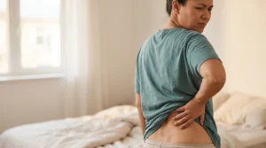
A small, rubbery lump near the top of the buttock can have several causes. One possibility is a fatty nodule sometimes called a back mouse. A clinician can examine a new or changing lump and decide whether it needs imaging.
Key takeaways
- Usually fat, not a disc. Most are fat pushed through a gap in the fascia; spinal discs sit far too deep to feel.
- One nickname, several labels. Two names often attached to it describe tender spots, not lumps.
- Pressure is the usual trigger. Waistbands, car-seat bolsters, hip belts and hard floors provoke a sore nodule more than bending does.
- Some lumps need a prompt check. Growth, a size over about 5 cm (2 inches), a hard or fixed feel, hot red skin or fever.
- Treatment is stepwise. Pressure management first; injection or removal for stubborn cases.
What are back mice, and what is their medical name?
The nickname describes mobile, rubbery nodules of fat in the lower back and upper buttock. Doctors usually call them episacroiliac or episacral lipomas, but the more accurate term is subfascial fat herniation: deeper fat that has bulged through a small gap in the fascia, the fibrous wrapping over the back muscles. A literature review in Pain Physician describes them as firm, mobile, rubbery lumps that may be fat squeezed out through the fascia.
| Name you may see | What it actually describes | Lump or tender spot? |
|---|---|---|
| Back mouse | 1990s lay nickname for mobile, tender nodules near the sacrum and pelvic rim | Lump |
| Episacroiliac or episacral lipoma | Medical label dating from the 1930s; most lack the capsule of a true lipoma | Lump |
| Subfascial or lumbar fascial fat herniation | Fat bulging through a fascial gap; the most accurate term | Lump |
| Fibro-fatty nodule, Copeman nodule | Older names for the same nodules | Lump |
| Iliac crest pain syndrome | Tenderness over the inner iliac crest, where ligaments and muscles attach | Tender spot |
| Multifidus triangle syndrome | 1950s label for recurrent low back pain traced to a tender area over the multifidus muscle low in the back | Tender spot |
The underlying fat pads are present even in very lean bodies, and older reports describe the nodules more often in women. A 1930s examination of 1,000 backs, cited in a 2021 historical review in Pain and Therapy, found nodules in about 30%, so having one is not a sign of trouble.
Where on the lower back do these lumps usually sit?

Most sit within a hand’s width of the two dimples at the base of the spine, either along the bony rim of the pelvis just above and outside each dimple or over the upper sacrum between them. They are often on both sides, one usually more tender, and range from pea-sized to walnut-sized or larger.
To map yours, stand with your hands on a counter and lean slightly forward, which makes nodules easier to feel:
- Find the dimples, which sit over the posterior superior iliac spines, the bony knobs at the back of the pelvis.
- Trace the iliac crest, the rim of the pelvis, up and out from each dimple, and note where the lump sits against it.
- Compare the other side. A hard knob mirrored on both sides is bone; a rubbery nodule that slides is not.
- Measure it once and write down the size and date, so growth becomes a fact rather than a worry.
Location also filters out look-alikes: a lump in the buttock crease near the tailbone suggests a pilonidal cyst, and a flank bulge that swells when you cough suggests a rare lumbar hernia. Dimples that ache with no lump have other causes, covered in a guide to why lower back dimples can hurt.
Why do some of these lumps hurt while others never do?
Pain seems to depend less on the nodule’s size than on what it presses against. One that slides freely in the fat layer can go unnoticed for years, while one trapped in a tight fascial gap, swollen with fluid or lying against a small skin nerve can become sharply tender, especially under outside pressure.
The thoracolumbar fascia is a diamond-shaped sheet of dense connective tissue over the low back, linking the latissimus dorsi to the gluteus maximus. It tightens like a drum skin whenever you bend, lift a toddler from a car seat or twist to reach the back seat, and small nerves pass through it via tiny openings. When deeper fat pushes up through one of those openings, it forms a small, mobile pocket, and biopsies have confirmed these nodules are fat. Plausible pain mechanisms include:
- Nerve contact with the superior cluneal nerves, small skin nerves that cross the pelvic rim to the buttock
- Trapped tension in swollen fat squeezed through a tight gap, like a finger stuck in a ring
- Outside pressure from a chair back, belt or floor pinning the nodule against bone

None of these is proven, and some clinicians argue that tender fat and back pain are simply both common. The practical test is whether pressing the nodule reproduces your familiar pain.
How can you tell a back mouse from other lumps?
For tenderness without a distinct lump, read about how lower back muscle tenderness can differ from a palpable lump.
A back mouse typically feels rubbery, slides a little under your fingertip, sits beneath the skin rather than in it, and is tender mainly when pressed. Lipomas are softer and painless, cysts are stuck to the skin, infections are red and hot, and anything hard, fixed, growing or larger than about 5 cm (2 inches) needs medical assessment.
| Lump | Feel and movement | Tender? | Usual spot | What it suggests |
|---|---|---|---|---|
| Back mouse | Firm, rubbery; slides but feels anchored | Often, when pressed | Near the dimples, pelvic rim, upper sacrum | Benign; manage pressure |
| Lipoma | Soft, doughy, moves freely | Usually not | Anywhere | Benign; remove if troublesome |
| Epidermoid (sebaceous) cyst | Smooth dome, may have a dark pore; moves with the skin | When inflamed | In the skin | Benign; do not squeeze |
| Abscess or infected cyst | Hot, swollen, fluid-filled | Very, even at rest | Anywhere; pilonidal type in the buttock crease | Seen within a day or two; same day with fever |
| Lumbar hernia | Soft bulge that grows with coughing | Variable | Flank, above the pelvic rim | Rare; surgical opinion |
| Muscle trigger point | Tender band within muscle, not a separate lump | Yes, with referred ache | Buttock, quadratus lumborum | Muscular; suits a physical therapist (physiotherapist) |
| Soft tissue sarcoma | Firm or hard, deep, often fixed | Often painless | Anywhere | Rare; prompt imaging |
Is it a slipped disc or sciatica instead?
A lump you can feel under the skin is not a disc. Lumbar discs sit several centimeters (a few inches) deep, in front of the spinal canal, and a herniation pushes back toward the nerves, not out to the skin. A sore fat nodule can still refer pain into the buttock or thigh, which is why the two get confused.
| Points to a nerve root | Points to the nodule |
|---|---|
| Leg pain worse than back pain, below the knee | Pain local or into the buttock and back of the thigh |
| Numbness or pins and needles in a strip down the leg | Pressing the lump reproduces your pain |
| Pain spikes with coughing or sneezing | Chair, belt or floor pressure matters more |
| Weakness, such as failing to rise onto tiptoe | Normal strength, sensation and reflexes |
A 2016 review in Pain Physician notes that these nodules can mimic disc herniation, and that the absence of neurological deficit helps tell them apart. If your symptoms fit the left column, compare them with what sciatica actually feels like; a bony landmark that aches with no lump may instead point to a problem in the sacroiliac joint itself.
When should a lump on your lower back be checked soon?
Arrange an examination for an unexplained lump, particularly if it grows, feels hard or fixed, or persists for two weeks. Get same-day medical advice if the skin is red or hot or you have fever. Get emergency care for new saddle numbness, new bladder or bowel changes, or severe or worsening weakness in both legs.
Quick check: is this lump something to get looked at soon?
- Has it grown since you measured it? Fat herniations change little, so growth is the strongest reason to scan.
- Is it wider than about 5 cm (2 inches), roughly the short edge of a bank card? Doctors usually image soft-tissue lumps above that size.
- Is it hard, or does it stay put when you try to slide it? Anchored lumps need imaging.
- Is the skin red, hot or draining, or do you feel feverish? That suggests infection.
- Night pain unrelated to lying on it, unexplained weight loss, or a past cancer? These call for ruling out a tumor.
Have a clinician assess a new, changing or persistent lump. Redness, heat or fever warrants same-day advice. The way a lump feels at home cannot establish whether it is harmless.
Will an ultrasound or MRI show the lump?
Ultrasound is the most useful scan for these nodules: it can show fat bulging between fascial layers, watch the nodule move, find the fascial gap, and help tell it apart from a cyst or solid mass. A standard lumbar spine MRI often says nothing about the lump because it is not looking for it, and X-rays will not show it.
Many people never need a scan: a small, mobile, unchanged lump that reproduces familiar pain is diagnosed by hand. Imaging earns its place for a warning feature, real doubt, or a planned injection or removal. Spine MRIs often miss the nodule because they center on discs and nerves and fat against fat gives little contrast; a 2021 historical review notes these nodules are generally not visible on routine X-ray or MRI.
- X-rays answer questions about bone, not fat; this guide to what a lower back X-ray can and cannot show explains the difference.
- If the lump itself is being scanned, ask for ultrasound first, with a skin marker over the spot.
- A dedicated soft-tissue MRI of that area, not a repeat spine MRI, is the next step if ultrasound shows more than simple fat.
Which common beliefs about these lumps are wrong?
The most common misconceptions are that the lump is a slipped disc you can feel, that a normal MRI proves it harmless, that firm massage can break it up, and that it might turn into cancer. Each leads to the wrong treatment or the wrong worry.
- “It is a disc that popped out.” Discs sit deep in the spine; this is in the fat under the skin.
- “A clear MRI means it cannot hurt.” Spine MRIs are not set up to assess small fat nodules.
- “Rolling it hard will dissolve it.” No evidence supports that; daily pressure keeps a sore nodule sore.
- “It could turn into cancer.” There is no evidence that these nodules transform.
From the Clinic: Dr. Arora’s Expert Insight
People tend to arrive with one finger pressed on the lump and, in the other hand, an MRI report that does not mention it at all. They have usually settled on one of two stories: the disc has come out and they can feel it, or the scan was thorough enough that the lump cannot matter. The first breeds a fear of bending; the second ends with the pain being called unexplained.
What I actually do is less dramatic: press the nodule and ask whether that is the pain or just a sore spot, press the mirror-image point, and check strength, sensation and reflexes. When the nodule reproduces the exact ache and the nerves test normal, the question becomes what keeps pressing on this one spot, and most people have never been asked it.
The failure I see most often comes from generic back pain programs built around lying on your back on a thin mat: knee hugs, bridges, pelvic tilts. For a sore fat herniation over the pelvis, each one parks body weight on the lump, turning a well-meant routine into a daily pressure test. Moving the same work onto a thicker mat with a gap under the nodule, or into side-lying, kneeling and standing versions, often settles things faster than new exercises would.
How do you calm a sore back mouse in the first 72 hours?
Take the pressure off the lump, stop prodding it, and keep moving. Most flares are driven by a belt, a seat or a floor pressing on the nodule, so changing contact points matters more than rest. Walking, side-lying and kneeling are fine; deep massage, rolling and repeated self-testing tend to keep it irritated.
What to do today
- Find the pressure source, whether a belt line, car-seat bolster, chair back, rucksack hip belt or exercise floor, and move it or pad around it.
- Make a gap, not a cushion: two folded towels either side of the lump offload it, while one cushion behind it presses harder.
- Stay on your feet: a few brief, gentle walks each day help more than resting on the sore spot. Sleep on your side.
- Use whichever of ice or heat suits your back for comfort; neither changes the lump.
- Measure it once, then leave it alone.
Still worse after 72 hours, or showing any quick-check warning sign? Get it examined.
A phased plan with clear progression points
Exercise will not remove a fat herniation. It eases guarding around a sore nodule, restores the bending people abandon when they fear a disc, and builds hip and back capacity. Progress on the criteria, not the calendar.
| Phase | What it includes, and why | Move on when |
|---|---|---|
| 1. Offload (days 1 to 7) | Walks; standing hip hinges, rebuilding bending with no contact on the lump; side-lying clamshells for the gluteus medius, which attaches just below the rim of the pelvis | Pressing the lump hurts only briefly; you can sit 30 minutes without a flare |
| 2. Restore (weeks 1 to 4) | Bird dog, with nothing touching the lower back; bridges on a thick mat with the towel gap, loading the gluteus maximus that blends into the fascia | No next-morning rise in tenderness after floor work |
| 3. Load (weeks 3 to 8) | Suitcase carries of 6 to 10 kg (13 to 22 lb), goblet squats and Romanian deadlifts, rehearsing real lifting | Every return checkpoint below passed without a next-day flare |

Do’s and don’ts while the nodule is tender
- Do set the car’s lumbar support above the nodule, using the seat setup for lower back pain while driving.
- Do shift a backpack hip belt just above or below a nodule on the iliac crest.
- Do keep bending and lifting; the lump is not a weakness in your spine.
- Don’t foam roll or squeeze it; a squeezed cyst can become infected.
- Don’t assume a changing lump is the harmless one you found last year.
Return-to-activity checkpoints
Each test is passed when pain settles within 30 minutes and the nodule is no more tender next morning:
- Sit or drive for 60 minutes, and lie on your back on a mat for 10 minutes.
- Romanian deadlift at half your usual weight for 3 sets of 8.
- Carry an 8 to 10 kg (18 to 22 lb) pack with the hip belt fastened for an hour.
- Run for 20 minutes with any running belt clear of the nodule.
What treatments help if the lump keeps hurting?
If a nodule is still clearly the pain source after several weeks of pressure management and graded activity, the next options are a physician’s injection of local anesthetic, sometimes with corticosteroid and repeated needling, or surgical removal with repair of the fascial gap. Both have encouraging reports, but the evidence comes mostly from small case series.
- Injection. It doubles as a test: if numbing the nodule abolishes your familiar pain, that supports it as the source. The 2016 Pain Physician review found anesthetic, with or without corticosteroid, plus repeated needling relieved pain in many case reports, yet the only randomized trial it identified, which used anesthetic without the repeated needling, found only mild, short-lived benefit. Ask whether ultrasound will guide the needle and whether a steroid, which can occasionally leave a small skin dent, is included.
- Surgical removal. Excision removes the fat pocket and closes the fascial defect. Older series reported good relief, but a 2021 review of that literature also describes recurrence: in one series, about 30% of nodules came back four to six months after removal. It suits a nodule confirmed as the pain source, for example by a convincing response to a numbing injection.
- Physical therapy. It cannot dissolve the lump, and an honest physical therapist (PT) will say so, but a PT can confirm the pattern, check the nerves, find your pressure sources and rebuild confidence with bending and lifting.
A warning about popping, cracking or breaking them up
Some practitioners and plenty of online videos promise to break up, pop or push back these nodules. There is no evidence that heavy pressure, scraping, cupping or manipulation removes a fat herniation, and repeated hard pressure can keep it inflamed.
The bigger risk is diagnostic: someone who treats every lump as a back mouse may be pressing on an infected cyst or a tumor. Walk away from anyone who offers to pierce or drain it outside a sterile medical setting, or works on a red, hot or growing lump without a medical opinion.
What to remember about back mice
A rubbery, mobile lump near your lower back dimples can have several causes. Arrange an examination if the lump is new, grows or persists. It often stays after the pain goes, so aim for a lump you stop noticing. Your action today: measure it, note the size and date, and remove whatever presses on it; growth, hardness, skin changes or fever mean an in-person examination.
Frequently asked questions
1. Can back mice go away on their own?
The pain often settles, but the lump usually stays. Once nothing presses on it, a nodule can become unnoticeable even though an examiner can still feel it.
2. Is it safe to massage a back mouse?
Light contact is harmless, but firm, repeated pressure on the nodule is a poor idea: it does not break up the fat and tends to keep it sore. Massage the muscles around it instead.
3. Could a lump on my lower back be cancer?
It is rarely cancer, but a new lump should not be diagnosed from a photo. Soft-tissue cancers here are uncommon and tend to be larger than about 5 cm (2 inches), deep, hard, fixed and growing; a small, rubbery, mobile nodule unchanged for months is far more likely to be benign fat.
4. What kind of doctor should check a lump on my lower back?
Start with your primary care doctor or GP, who can examine it and arrange an ultrasound. Pain or musculoskeletal physicians usually give injections and general or plastic surgeons remove persistent nodules; a physical therapist or PT can address pressure triggers and refer you on.
5. Are these lumps related to lower back dimples?
Not directly, but they share an address. The dimples mark the posterior superior iliac spines, and fat nodules often sit within a hand’s width of them; a dimple that aches with no lump points to the joint and ligaments beneath instead.
6. Can I keep running, lifting or hiking with a painful lump on my back?
Yes, in most cases, because the lump does not weaken the spine. Change contact pressure from belts, hip straps and benches rather than stopping the activity, and scale back only if the nodule is more tender the next morning.
7. Will losing weight make the lumps disappear?
Probably not, and there is no good evidence that it does. The fat pads involved occur even in very lean people, and the underlying problem is a fascial gap rather than total body fat.
Medical disclaimer
This page explains a common, usually benign cause of lower back lumps; it cannot identify yours. Calling a lump a fat nodule requires an in-person examination, sometimes ultrasound, and ruling out cysts, infection and rarer tumors. A growing, hard or fixed lump, or one with fever, skin changes, night pain or weight loss, needs a doctor, and injection or surgery decisions need a risk discussion with the clinician involved.
References
- Bicket MC, Simmons C, Zheng Y. The best-laid plans of “back mice” and men: a case report and literature review of episacroiliac lipoma. Pain Physician. 2016;19(3):181-188. PMID: 27008292.
- Cañis Parera M, Expósito Izquierdo M, Cabré Vila JJ. Historical review of studies on sacroiliac fatty nodules (recently termed “back mice”) as a potential cause of low back pain. Pain Ther. 2021;10(2):1029-1050. doi:10.1007/s40122-021-00321-5. PMID: 34528160.