If you’ve recently had an MRI for your back pain, you might have seen the term “Modic changes on MRI” in the report. This finding can be confusing, but understanding what it means is a crucial step in unraveling the mystery of your discomfort. Far from just medical jargon, Modic changes on MRI offer important clues about the health of your spine and point directly to a specific source of chronic discomfort: vertebrogenic low back pain.
What Exactly Are Modic Changes?
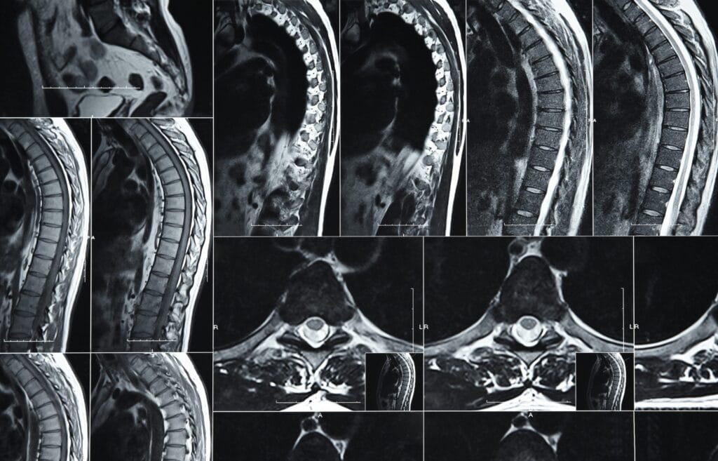
To put it simply, Modic changes are alterations visible on an MRI scan that occur in the bone marrow of your vertebrae (the individual bones of your spine) and the adjacent vertebral endplates. These endplates are the surfaces where your vertebrae connect with your intervertebral discs.
When these areas experience stress, inflammation, or degeneration, they undergo changes that show up distinctly on an MRI, signaling a process happening within the bone itself. They are named after Dr. Michael Modic, who first described them.
The Three Types of Modic Changes
Modic changes are typically classified into three types, each with a different appearance on MRI and potentially different implications:
Type 1 Modic Changes: The “Active” Stage
- Appearance on MRI: Bright on T2-weighted images and dark on T1-weighted images.
- What it means: This type indicates bone marrow edema and inflammation. Think of it like active swelling and irritation within the bone. Type 1 changes are often strongly associated with pain, making them a key focus when discussing Modic changes on MRI in relation to discomfort. They suggest a more acute or active inflammatory process, commonly seen in cases of vertebrogenic low back pain.
Type 2 Modic Changes: The “Fatty” Stage
- Appearance on MRI: Bright on both T1- and T2-weighted images.
- What it means: This type signifies fatty degeneration of the bone marrow. It often represents a more chronic or healed stage where the active inflammation has subsided and been replaced by fat. Type 2 changes can sometimes be a progression from Type 1, and while they can be associated with pain, they are generally considered less painful than Type 1.
Type 3 Modic Changes: The “Sclerotic” Stage
- Appearance on MRI: Dark on both T1- and T2-weighted images.
- What it means: This type indicates sclerosis or hardening of the bone. It’s the least common type and typically suggests long-standing degenerative changes where the bone has become dense and fibrous. Type 3 changes are usually not directly associated with active pain.
Why Do Modic Changes Occur?
Modic changes on MRI are generally a response to stress and degeneration at the vertebral endplates. Common causes and associations include:
- Degenerative Disc Disease: As spinal discs lose height and hydration, they put more stress on the endplates, leading to inflammation and breakdown.
- Microtrauma: Small, repetitive injuries to the endplates can trigger an inflammatory response.
- Infection: Though less common, infections can also cause changes similar to Modic Type 1.
- Biomechanical Stress: Poor posture, heavy lifting, and prolonged sitting can contribute to the stress on these areas, often seen in the development of vertebrogenic low back pain.
The Connection Between Modic Changes and Pain
Not everyone with Modic changes on MRI experiences back pain, and not all back pain is caused by Modic changes. However, there’s a strong and growing body of research linking Type 1 Modic changes on MRI specifically to chronic low back pain, particularly vertebrogenic low back pain.
When Type 1 changes are present, it suggests active inflammation within the vertebral bone, where tiny nerves (like the basivertebral nerve) can be irritated, sending pain signals. This is why understanding Modic changes on MRI and their link to specific conditions like vertebrogenic low back pain can be a game-changer for many individuals whose discomfort hasn’t responded to traditional treatments.
Diagnosing Modic Changes: What to Expect
If your doctor suspects an issue with your vertebral endplates, an MRI scan is the primary diagnostic tool. Modic changes on MRI are clearly visible, making it an invaluable part of the diagnostic process for back pain.
While X-rays show bone structure, they don’t reveal the subtle changes in bone marrow that an MRI can. A detailed consultation with your spine specialist, combined with your MRI findings, helps determine if these specific Modic changes on MRI are indeed contributing to your pain and if it points towards vertebrogenic low back pain.
What Happens After a Modic Change Diagnosis?
A diagnosis of Modic changes on MRI doesn’t automatically mean surgery or a specific treatment. The approach typically depends on the type of Modic change, the severity of your pain, and how it impacts your daily life.
- Conservative Management: This often includes physiotherapy, core strengthening, anti-inflammatory medications, and lifestyle adjustments, similar to initial approaches for vertebrogenic low back pain.
- Advanced Interventions: For persistent pain associated with Type 1 Modic changes, advanced procedures like basivertebral nerve ablation (BVNA) may be considered. This procedure specifically targets the nerves involved in sending pain signals from the inflamed endplates, a common treatment for vertebrogenic low back pain.
Living with Modic Changes
Understanding your MRI findings can be empowering. Knowing that your back pain might have a specific, identifiable cause like Modic changes on MRI can lead to more targeted and effective treatment strategies for conditions like vertebrogenic low back pain.
It’s crucial to work closely with your healthcare provider to develop a personalized management plan. Maintaining good posture, engaging in regular appropriate exercise, and managing your weight are all vital steps in supporting overall spinal health and potentially alleviating pain associated with these changes.
FAQs about Modic Changes
Are Modic changes always painful? No, particularly Type 2 and Type 3 changes may be asymptomatic. Type 1 Modic changes on MRI are most strongly associated with pain and are often linked to vertebrogenic low back pain.
Can Modic changes disappear? Type 1 Modic changes can sometimes convert to Type 2, indicating a reduction in active inflammation. They generally don’t disappear completely from the MRI but can become less symptomatic.
Do Modic changes require surgery? Not necessarily. Many cases are managed successfully with conservative or minimally invasive treatments. Surgery is typically a last resort, as discussed in treatments for vertebrogenic low back pain.
Is there a specific exercise for Modic changes? No single exercise targets Modic changes on MRI directly, but core strengthening, improving posture, and exercises that promote spinal stability can help manage associated pain. Always consult a physical therapist.
How common are Modic changes? They are quite common, especially in people with chronic low back pain and degenerative disc disease, with prevalence varying depending on the study population. Their presence often prompts investigation for vertebrogenic low back pain.
With 20+ years of clinical experience in spine, joint, and pain management, Dr. Vivek ensures every article reflects accurate, trustworthy, and up-to-date health advice.
Learn more about Dr. Vivek’s qualifications & journey →